 Movie
Movie Controller

[English] 日本語
 Yorodumi
- PDB-1mmn: X-RAY STRUCTURES OF THE MGADP, MGATPGAMMAS, AND MGAMPPNP COMPLEXE... -
+ Open data

- Basic information
| Entry | Database: PDB / ID: 1mmn | ||||||
|---|---|---|---|---|---|---|---|
| Title | X-RAY STRUCTURES OF THE MGADP, MGATPGAMMAS, AND MGAMPPNP COMPLEXES OF THE DICTYOSTELIUM DISCOIDEUM MYOSIN MOTOR DOMAIN | ||||||
 Components Components | MYOSIN | ||||||
 Keywords Keywords | COILED COIL / MYOSIN / DICTYOSTELIUM / MOTOR / NUCLEOTIDE ANALOGUES / AMPPNP / ATPASE / ACTIN-BINDING | ||||||
| Function / homology |  Function and homology information Function and homology informationuropod retraction / cytoplasmic actin-based contraction involved in forward cell motility / phagocytic cup base / pathogen-containing vacuole / response to differentiation-inducing factor 1 / equatorial cell cortex / contractile actin filament bundle assembly / pseudopodium retraction / cell trailing edge / contractile vacuole organization ...uropod retraction / cytoplasmic actin-based contraction involved in forward cell motility / phagocytic cup base / pathogen-containing vacuole / response to differentiation-inducing factor 1 / equatorial cell cortex / contractile actin filament bundle assembly / pseudopodium retraction / cell trailing edge / contractile vacuole organization / myosin filament assembly / aggregation involved in sorocarp development / culmination involved in sorocarp development / adenyl nucleotide binding / RHO GTPases activate PAKs / calcium-dependent ATPase activity / actomyosin contractile ring / hypotonic response / uropod / apical cortex / negative regulation of actin filament polymerization / actin-myosin filament sliding / bleb assembly / detection of mechanical stimulus / substrate-dependent cell migration, cell extension / actomyosin / filopodium assembly / early phagosome / myosin filament / cortical actin cytoskeleton organization / myosin II complex / cortical actin cytoskeleton / microfilament motor activity / cleavage furrow / pseudopodium / mitotic cytokinesis / cytoskeletal motor activity / response to cAMP / response to mechanical stimulus / 14-3-3 protein binding / cell motility / response to hydrogen peroxide / chemotaxis / actin filament binding / intracellular protein localization / regulation of cell shape / extracellular matrix / cytoplasmic vesicle / cell cortex / cytoskeleton / calmodulin binding / ATP binding / identical protein binding / cytosol / cytoplasm Similarity search - Function | ||||||
| Biological species |  | ||||||
| Method |  X-RAY DIFFRACTION / SYNCHROTRON / MOLECULAR REPLACEMENT / Resolution: 2.1 Å X-RAY DIFFRACTION / SYNCHROTRON / MOLECULAR REPLACEMENT / Resolution: 2.1 Å | ||||||
 Authors Authors | Gulick, A.M. / Bauer, C.B. / Thoden, J.B. / Rayment, I. | ||||||
 Citation Citation | Journal: Biochemistry / Year: 1997 Title: X-ray structures of the MgADP, MgATPgammaS, and MgAMPPNP complexes of the Dictyostelium discoideum myosin motor domain. Authors: Gulick, A.M. / Bauer, C.B. / Thoden, J.B. / Rayment, I. #1: Journal: Biochemistry / Year: 1995Title: X-Ray Structures of the Myosin Motor Domain of Dictyostelium Discoideum Complexed with Mgadp(Dot)Befx and Mgadp(Dot)Alf4- Authors: Fisher, A.J. / Smith, C.A. / Thoden, J.B. / Smith, R. / Sutoh, K. / Holden, H.M. / Rayment, I. | ||||||
| History |
|
- Structure visualization
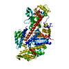
| Structure viewer | Molecule: MolmilJmol/JSmol |
|---|
- Downloads & links
-Download
| PDBx/mmCIF format | 1mmn.cif.gz | 172.3 KB | Display | PDBx/mmCIF format |
|---|---|---|---|---|
| PDB format | pdb1mmn.ent.gz | 131.7 KB | Display | PDB format |
| PDBx/mmJSON format | 1mmn.json.gz | Tree view | PDBx/mmJSON format | |
| Others |  Other downloads Other downloads |
-Validation report
| Arichive directory | https://data.pdbj.org/pub/pdb/validation_reports/mm/1mmnftp://data.pdbj.org/pub/pdb/validation_reports/mm/1mmn | HTTPS FTP |
|---|
-Related structure data
| Related structure data | 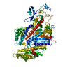 1mmaC 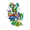 1mmgC 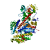 1mmdS S: Starting model for refinement C: citing same article ( |
|---|---|
| Similar structure data |


-Links
 PDBj
PDBj


- Assembly
| Deposited unit | 
| ||||||||
|---|---|---|---|---|---|---|---|---|---|
| 1 |
| ||||||||
| Unit cell |
|
-Components
| #1: Protein | Mass: 86781.094 Da / Num. of mol.: 1 / Fragment: MOTOR DOMAIN / Mutation: Q760L, R761P, I762N Source method: isolated from a genetically manipulated source Details: GENETICALLY TRUNCATED HEAD OF MYOSIN FROM DICTYOSTELIUM. LIGANDS MG2+, AMPPNP Source: (gene. exp.) |
|---|---|
| #2: Chemical | ChemComp-MG /   Mass: 24.305 Da / Num. of mol.: 1 / Source method: obtained synthetically / Formula: Mg Mass: 24.305 Da / Num. of mol.: 1 / Source method: obtained synthetically / Formula: Mg |
| #3: Chemical | ChemComp-ANP /   Mass: 506.196 Da / Num. of mol.: 1 / Source method: obtained synthetically / Formula: C10H17N6O12P3 / Comment: AMP-PNP, energy-carrying molecule analogue*YM Mass: 506.196 Da / Num. of mol.: 1 / Source method: obtained synthetically / Formula: C10H17N6O12P3 / Comment: AMP-PNP, energy-carrying molecule analogue*YM |
| #4: Water | ChemComp-HOH /  Mass: 18.015 Da / Num. of mol.: 477 / Source method: isolated from a natural source / Formula: H2O Mass: 18.015 Da / Num. of mol.: 477 / Source method: isolated from a natural source / Formula: H2O |
-Experimental details
-Experiment
| Experiment | Method: X-RAY DIFFRACTION / Number of used crystals: 1 |
|---|
- Sample preparation
| Crystal | Density Matthews: 2.92 Å3/Da / Density % sol: 57.81 % | ||||||||||||||||||||||||||||||||||||||||||||||||
|---|---|---|---|---|---|---|---|---|---|---|---|---|---|---|---|---|---|---|---|---|---|---|---|---|---|---|---|---|---|---|---|---|---|---|---|---|---|---|---|---|---|---|---|---|---|---|---|---|---|
| Crystal grow | pH: 7 / Details: pH 7.0 | ||||||||||||||||||||||||||||||||||||||||||||||||
| Crystal grow | *PLUS Temperature: 4 ℃ / Method: batch method | ||||||||||||||||||||||||||||||||||||||||||||||||
| Components of the solutions | *PLUS
|
-Data collection
| Diffraction | Mean temperature: 113 K |
|---|---|
| Diffraction source | Source: SYNCHROTRON / Site: CHESS  / Beamline: F1 / Wavelength: 0.908 / Beamline: F1 / Wavelength: 0.908 |
| Detector | Date: May 1, 1996 |
| Radiation | Monochromatic (M) / Laue (L): M / Scattering type: x-ray |
| Radiation wavelength | Wavelength: 0.908 Å / Relative weight: 1 |
| Reflection | Resolution: 2.1→20 Å / Num. obs: 58467 / % possible obs: 92.6 % / Redundancy: 4.2 % / Rmerge(I) obs: 0.075 / Net I/σ(I): 14 |
| Reflection shell | Resolution: 2.1→2.18 Å / % possible all: 55.8 |
| Reflection | *PLUS Num. measured all: 244680 |
| Reflection shell | *PLUS % possible obs: 55.8 % |
- Processing
| Software |
| ||||||||||||||||||||||||||||||
|---|---|---|---|---|---|---|---|---|---|---|---|---|---|---|---|---|---|---|---|---|---|---|---|---|---|---|---|---|---|---|---|
| Refinement | Method to determine structure: MOLECULAR REPLACEMENT Starting model: PDB ENTRY 1MMD Resolution: 2.1→30 Å / σ(F): 0
| ||||||||||||||||||||||||||||||
| Refinement step | Cycle: LAST / Resolution: 2.1→30 Å
| ||||||||||||||||||||||||||||||
| Refine LS restraints |
| ||||||||||||||||||||||||||||||
| Software | *PLUS Name: TNT / Classification: refinement | ||||||||||||||||||||||||||||||
| Refinement | *PLUS Rfactor obs: 0.198 | ||||||||||||||||||||||||||||||
| Solvent computation | *PLUS | ||||||||||||||||||||||||||||||
| Displacement parameters | *PLUS Biso mean: 37.4 Å2 | ||||||||||||||||||||||||||||||
| Refine LS restraints | *PLUS Type: t_plane_restr / Dev ideal: 0.006 |
