 Movie
Movie Controller

[English] 日本語
 Yorodumi
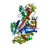
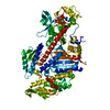
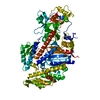
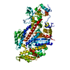
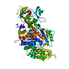
- PDB-1d1c: DICTYOSTELIUM MYOSIN S1DC (MOTOR DOMAIN FRAGMENT) COMPLEXED WITH ... -
+ Open data

- Basic information
| Entry | Database: PDB / ID: 1d1c | ||||||
|---|---|---|---|---|---|---|---|
| Title | DICTYOSTELIUM MYOSIN S1DC (MOTOR DOMAIN FRAGMENT) COMPLEXED WITH N-METHYL-O-NITROPHENYL AMINOETHYLDIPHOSPHATE BERYLLIUM TRIFLUORIDE. | ||||||
 Components Components | MYOSIN | ||||||
 Keywords Keywords | CONTRACTILE PROTEIN / MYOSIN / MOTILITY / ACTIN-BINDING / MOTOR DOMAIN / NANOLOGS | ||||||
| Function / homology |  Function and homology information Function and homology informationuropod retraction / cytoplasmic actin-based contraction involved in forward cell motility / phagocytic cup base / pathogen-containing vacuole / response to differentiation-inducing factor 1 / equatorial cell cortex / contractile actin filament bundle assembly / pseudopodium retraction / cell trailing edge / contractile vacuole organization ...uropod retraction / cytoplasmic actin-based contraction involved in forward cell motility / phagocytic cup base / pathogen-containing vacuole / response to differentiation-inducing factor 1 / equatorial cell cortex / contractile actin filament bundle assembly / pseudopodium retraction / cell trailing edge / contractile vacuole organization / myosin filament assembly / aggregation involved in sorocarp development / culmination involved in sorocarp development / adenyl nucleotide binding / RHO GTPases activate PAKs / calcium-dependent ATPase activity / hypotonic response / actomyosin contractile ring / uropod / apical cortex / negative regulation of actin filament polymerization / actin-myosin filament sliding / detection of mechanical stimulus / substrate-dependent cell migration, cell extension / bleb assembly / actomyosin / filopodium assembly / myosin filament / early phagosome / cortical actin cytoskeleton organization / myosin II complex / cortical actin cytoskeleton / microfilament motor activity / cleavage furrow / pseudopodium / mitotic cytokinesis / cytoskeletal motor activity / response to cAMP / response to mechanical stimulus / 14-3-3 protein binding / response to hydrogen peroxide / cell motility / chemotaxis / actin filament binding / intracellular protein localization / regulation of cell shape / extracellular matrix / cytoplasmic vesicle / cell cortex / cytoskeleton / calmodulin binding / ATP binding / identical protein binding / cytoplasm / cytosol Similarity search - Function | ||||||
| Biological species |  | ||||||
| Method |  X-RAY DIFFRACTION / SYNCHROTRON / Resolution: 2.3 Å X-RAY DIFFRACTION / SYNCHROTRON / Resolution: 2.3 Å | ||||||
 Authors Authors | Gulick, A.M. / Bauer, C.B. / Thoden, J.B. / Pate, E. / Yount, R.G. / Rayment, I. | ||||||
 Citation Citation | Journal: J.Biol.Chem. / Year: 2000 Title: X-ray structures of the Dictyostelium discoideum myosin motor domain with six non-nucleotide analogs. Authors: Gulick, A.M. / Bauer, C.B. / Thoden, J.B. / Pate, E. / Yount, R.G. / Rayment, I. | ||||||
| History |
|
- Structure visualization
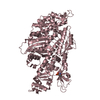
| Structure viewer | Molecule: MolmilJmol/JSmol |
|---|
- Downloads & links
-Download
| PDBx/mmCIF format | 1d1c.cif.gz | 173.8 KB | Display | PDBx/mmCIF format |
|---|---|---|---|---|
| PDB format | pdb1d1c.ent.gz | 136 KB | Display | PDB format |
| PDBx/mmJSON format | 1d1c.json.gz | Tree view | PDBx/mmJSON format | |
| Others |  Other downloads Other downloads |
-Validation report
| Arichive directory | https://data.pdbj.org/pub/pdb/validation_reports/d1/1d1cftp://data.pdbj.org/pub/pdb/validation_reports/d1/1d1c | HTTPS FTP |
|---|
-Related structure data


-Links
 PDBj
PDBj


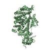
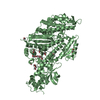
- Assembly
| Deposited unit | 
| ||||||||
|---|---|---|---|---|---|---|---|---|---|
| 1 |
| ||||||||
| Unit cell |
|
-Components
| #1: Protein | Mass: 86735.109 Da / Num. of mol.: 1 / Fragment: S1DC MOTOR DOMAIN / Mutation: Q760P, R761N Source method: isolated from a genetically manipulated source Source: (gene. exp.) |
|---|---|
| #2: Chemical | ChemComp-MG /   Mass: 24.305 Da / Num. of mol.: 1 / Source method: obtained synthetically / Formula: Mg Mass: 24.305 Da / Num. of mol.: 1 / Source method: obtained synthetically / Formula: Mg |
| #3: Chemical | ChemComp-NMQ / 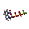  Mass: 421.162 Da / Num. of mol.: 1 / Source method: obtained synthetically / Formula: C9H13BeF3N2O9P2 Mass: 421.162 Da / Num. of mol.: 1 / Source method: obtained synthetically / Formula: C9H13BeF3N2O9P2 |
| #4: Water | ChemComp-HOH /  Mass: 18.015 Da / Num. of mol.: 591 / Source method: isolated from a natural source / Formula: H2O Mass: 18.015 Da / Num. of mol.: 591 / Source method: isolated from a natural source / Formula: H2O |
-Experimental details
-Experiment
| Experiment | Method: X-RAY DIFFRACTION / Number of used crystals: 1 |
|---|
- Sample preparation
| Crystal | Density Matthews: 2.93 Å3/Da / Density % sol: 58.02 % | ||||||||||||||||||||||||||||||||||||
|---|---|---|---|---|---|---|---|---|---|---|---|---|---|---|---|---|---|---|---|---|---|---|---|---|---|---|---|---|---|---|---|---|---|---|---|---|---|
| Crystal grow | Temperature: 277 K / Method: microbatch / pH: 7 Details: 8.3 % PEG 8000 125 MM NACL 3 MM DTT, pH 7.0, MICROBATCH, temperature 4.0K | ||||||||||||||||||||||||||||||||||||
| Crystal grow | *PLUS Method: batch method | ||||||||||||||||||||||||||||||||||||
| Components of the solutions | *PLUS
|
-Data collection
| Diffraction | Mean temperature: 100 K |
|---|---|
| Diffraction source | Source: SYNCHROTRON / Site: SSRL  / Beamline: BL7-1 / Wavelength: 0.908 / Beamline: BL7-1 / Wavelength: 0.908 |
| Detector | Type: MARRESEARCH / Detector: IMAGE PLATE / Date: Aug 7, 1996 |
| Radiation | Protocol: SINGLE WAVELENGTH / Monochromatic (M) / Laue (L): M / Scattering type: x-ray |
| Radiation wavelength | Wavelength: 0.908 Å / Relative weight: 1 |
| Reflection | Resolution: 2→25 Å / Num. all: 42098 / Num. obs: 42098 / % possible obs: 90.9 % / Observed criterion σ(F): 0 / Observed criterion σ(I): -3 / Redundancy: 3.16 % / Rmerge(I) obs: 0.075 / Net I/σ(I): 4.9 |
| Reflection shell | Resolution: 2.3→2.4 Å / Redundancy: 2 % / Rmerge(I) obs: 0.243 / % possible all: 93 |
| Reflection | *PLUS Num. all: 46312 / Num. measured all: 133036 |
| Reflection shell | *PLUS % possible obs: 93 % |
- Processing
| Software |
| ||||||||||||||||||||||||||||||
|---|---|---|---|---|---|---|---|---|---|---|---|---|---|---|---|---|---|---|---|---|---|---|---|---|---|---|---|---|---|---|---|
| Refinement | Resolution: 2.3→25 Å / σ(F): 0 / σ(I): 0 / Stereochemistry target values: ENGH & HUBER
| ||||||||||||||||||||||||||||||
| Refinement step | Cycle: LAST / Resolution: 2.3→25 Å
| ||||||||||||||||||||||||||||||
| Refine LS restraints |
| ||||||||||||||||||||||||||||||
| Software | *PLUS Name: TNT / Version: 5E / Classification: refinement | ||||||||||||||||||||||||||||||
| Refine LS restraints | *PLUS Type: t_plane_restr / Dev ideal: 0.013 |
