 Movie
Movie Controller

[English] 日本語
 Yorodumi
- PDB-3zzp: Circular permutant of ribosomal protein S6, lacking edge strand b... -
+ Open data

- Basic information
| Entry | Database: PDB / ID: 3zzp | ||||||
|---|---|---|---|---|---|---|---|
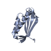
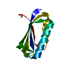
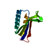
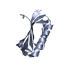
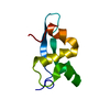
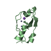
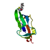
| Title | Circular permutant of ribosomal protein S6, lacking edge strand beta- 2 of wild-type S6. | ||||||
 Components Components | RIBOSOMAL PROTEIN S6 | ||||||
 Keywords Keywords | RIBOSOMAL PROTEIN / PROTEIN FOLDING / RNA-BINDING | ||||||
| Function / homology |  Function and homology information Function and homology informationsmall ribosomal subunit rRNA binding / structural constituent of ribosome / ribosome / translation / ribonucleoprotein complex / cytoplasm Similarity search - Function | ||||||
| Biological species |   THERMUS THERMOPHILUS (bacteria) THERMUS THERMOPHILUS (bacteria) | ||||||
| Method |  X-RAY DIFFRACTION / SYNCHROTRON / MOLECULAR REPLACEMENT / Resolution: 0.96 Å X-RAY DIFFRACTION / SYNCHROTRON / MOLECULAR REPLACEMENT / Resolution: 0.96 Å | ||||||
 Authors Authors | Saraboji, K. / Haglund, E. / Lindberg, M.O. / Oliveberg, M. / Logan, D.T. | ||||||
 Citation Citation | Journal: J.Biol.Chem. / Year: 2012 Title: Trimming Down a Protein Structure to its Bare Foldons: Spatial Organization of the Cooperative Unit. Authors: Haglund, E. / Danielsson, J. / Kadhirvel, S. / Lindberg, M.O. / Logan, D.T. / Oliveberg, M. | ||||||
| History |
|
- Structure visualization
| Structure viewer | Molecule: MolmilJmol/JSmol |
|---|
- Downloads & links
-Download
| PDBx/mmCIF format | 3zzp.cif.gz | 49.9 KB | Display | PDBx/mmCIF format |
|---|---|---|---|---|
| PDB format | pdb3zzp.ent.gz | 34.8 KB | Display | PDB format |
| PDBx/mmJSON format | 3zzp.json.gz | Tree view | PDBx/mmJSON format | |
| Others |  Other downloads Other downloads |
-Validation report
| Summary document | 3zzp_validation.pdf.gz | 420 KB | Display | wwPDB validaton report |
|---|---|---|---|---|
| Full document | 3zzp_full_validation.pdf.gz | 422.2 KB | Display | |
| Data in XML | 3zzp_validation.xml.gz | 7.1 KB | Display | |
| Data in CIF | 3zzp_validation.cif.gz | 9.5 KB | Display | |
| Arichive directory | https://data.pdbj.org/pub/pdb/validation_reports/zz/3zzpftp://data.pdbj.org/pub/pdb/validation_reports/zz/3zzp | HTTPS FTP |
-Related structure data
| Related structure data |  1risS S: Starting model for refinement |
|---|---|
| Similar structure data |


-Links
 PDBj
PDBj


- Assembly
| Deposited unit | 
| ||||||||
|---|---|---|---|---|---|---|---|---|---|
| 1 |
| ||||||||
| Unit cell |
|
-Components
| #1: Protein | Mass: 9031.086 Da / Num. of mol.: 1 / Fragment: RESIDUES 3-35,55-93 Source method: isolated from a genetically manipulated source Details: CIRCULAR PERMUTANT OF THE RIBOSOMAL PROTEIN S6 FROM THERMUS THERMOPHILUS, WHICH HAS A LINKER BETWEEN THE WILD-TYPE N-AND C-TERMINI AND AN INCISION BETWEEN K54 AND D55 WITHOUT THE EDGE STRAND ...Details: CIRCULAR PERMUTANT OF THE RIBOSOMAL PROTEIN S6 FROM THERMUS THERMOPHILUS, WHICH HAS A LINKER BETWEEN THE WILD-TYPE N-AND C-TERMINI AND AN INCISION BETWEEN K54 AND D55 WITHOUT THE EDGE STRAND BETA-2 OF WILD-TYPE S6. (RESIDUES 36-54). Source: (gene. exp.) THERMUS THERMOPHILUS (bacteria) / Production host: |
|---|---|
| #2: Water | ChemComp-HOH /  Mass: 18.015 Da / Num. of mol.: 129 / Source method: isolated from a natural source / Formula: H2O Mass: 18.015 Da / Num. of mol.: 129 / Source method: isolated from a natural source / Formula: H2O |
| Sequence details | CIRCULAR PERMUTANT OF THE RIBOSOMAL PROTEIN S6, WHICH HAS A LINKER BETWEEN THE WILD-TYPE N- AND C- ...CIRCULAR PERMUTANT OF THE RIBOSOMAL PROTEIN S6, WHICH HAS A LINKER BETWEEN THE WILD-TYPE N- AND C-TERMINI AND AN INCISION BETWEEN K54 AND D55 WITHOUT THE STRAND BETA-2 OF WILD-TYPE S6 (RESIDUES 36-54). |
-Experimental details
-Experiment
| Experiment | Method: X-RAY DIFFRACTION / Number of used crystals: 1 |
|---|
- Sample preparation
| Crystal | Density Matthews: 1.56 Å3/Da / Density % sol: 20.9 % / Description: NONE |
|---|---|
| Crystal grow | pH: 6.5 Details: 1.9M AMMONIUM SULPHATE, 0.1M MES PH 6.5, 8% 1,4 DIOXANE |
-Data collection
| Diffraction | Mean temperature: 100 K |
|---|---|
| Diffraction source | Source: SYNCHROTRON / Site: MAX II  / Beamline: I911-2 / Wavelength: 1.038 / Beamline: I911-2 / Wavelength: 1.038 |
| Detector | Type: MARRESEARCH / Detector: CCD / Date: Mar 13, 2009 |
| Radiation | Monochromator: BENT SI (111) CRYSTAL / Protocol: SINGLE WAVELENGTH / Monochromatic (M) / Laue (L): M / Scattering type: x-ray |
| Radiation wavelength | Wavelength: 1.038 Å / Relative weight: 1 |
| Reflection | Resolution: 0.96→20 Å / Num. obs: 38300 / % possible obs: 99.3 % / Observed criterion σ(I): 0 / Redundancy: 6 % / Rmerge(I) obs: 0.06 / Net I/σ(I): 16 |
| Reflection shell | Resolution: 0.96→0.99 Å / Redundancy: 4.86 % / Rmerge(I) obs: 0.65 / Mean I/σ(I) obs: 3 / % possible all: 98.2 |
- Processing
| Software |
| |||||||||||||||||||||||||||||||||
|---|---|---|---|---|---|---|---|---|---|---|---|---|---|---|---|---|---|---|---|---|---|---|---|---|---|---|---|---|---|---|---|---|---|---|
| Refinement | Method to determine structure: MOLECULAR REPLACEMENT Starting model: PDB ENTRY 1RIS Resolution: 0.96→20 Å / Num. parameters: 6866 / Num. restraintsaints: 8147 / Cross valid method: FREE R-VALUE / σ(F): 0 / Stereochemistry target values: ENGH AND HUBER
| |||||||||||||||||||||||||||||||||
| Refine analyze | Num. disordered residues: 9 / Occupancy sum hydrogen: 542.32 / Occupancy sum non hydrogen: 733.74 | |||||||||||||||||||||||||||||||||
| Refinement step | Cycle: LAST / Resolution: 0.96→20 Å
| |||||||||||||||||||||||||||||||||
| Refine LS restraints |
|
