 Movie
Movie Controller

[English] 日本語
 Yorodumi
- PDB-1qjh: Protein Aggregation and Alzheimer's Disease: Crystallographic Ana... -
+ Open data

- Basic information
| Entry | Database: PDB / ID: 1qjh | ||||||
|---|---|---|---|---|---|---|---|
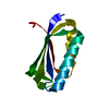
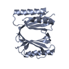
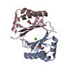
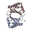
| Title | Protein Aggregation and Alzheimer's Disease: Crystallographic Analysis of the Phenomenon. Engineered version of the ribosomal protein S6 used as a stable scaffold to study oligomerization. | ||||||
 Components Components | 30S ribosomal protein S6 | ||||||
 Keywords Keywords | RIBOSOMAL PROTEIN / ALZHEIMER DISEASE / RIBOSOMAL PROTEIN S6 / OLIGOMERIZATION | ||||||
| Function / homology |  Function and homology information Function and homology informationsmall ribosomal subunit rRNA binding / structural constituent of ribosome / ribosome / translation / ribonucleoprotein complex / cytoplasm Similarity search - Function | ||||||
| Biological species |   Thermus thermophilus (bacteria) Thermus thermophilus (bacteria) | ||||||
| Method |  X-RAY DIFFRACTION / SYNCHROTRON / MOLECULAR REPLACEMENT / Resolution: 2.2 Å X-RAY DIFFRACTION / SYNCHROTRON / MOLECULAR REPLACEMENT / Resolution: 2.2 Å | ||||||
 Authors Authors | Kristensen, O. / Otzen, D.E. / Oliveberg, M. | ||||||
 Citation Citation | Journal: Proc. Natl. Acad. Sci. U.S.A. / Year: 2000 Title: Designed protein tetramer zipped together with a hydrophobic Alzheimer homology: a structural clue to amyloid assembly. Authors: Otzen, D.E. / Kristensen, O. / Oliveberg, M. #1: Journal: Embo J. / Year: 1994Title: Crystal Structure of the Ribosomal Protein S6 from Thermus Thermophilus Authors: Lindahl, M. / Svensson, L.A. / Liljas, A. / Sedelnikova, S.E. / Eliseikina, I.A. / Fomenkova, N.P. / Nevskaya, N. / Nikonov, S.V. / Garber, M.B. / Muranova, T.A. / Rykonova, A.I. / Amons, R. | ||||||
| History |
|
- Structure visualization
| Structure viewer | Molecule: MolmilJmol/JSmol |
|---|
- Downloads & links
-Download
| PDBx/mmCIF format | 1qjh.cif.gz | 33 KB | Display | PDBx/mmCIF format |
|---|---|---|---|---|
| PDB format | pdb1qjh.ent.gz | 21.8 KB | Display | PDB format |
| PDBx/mmJSON format | 1qjh.json.gz | Tree view | PDBx/mmJSON format | |
| Others |  Other downloads Other downloads |
-Validation report
| Arichive directory | https://data.pdbj.org/pub/pdb/validation_reports/qj/1qjhftp://data.pdbj.org/pub/pdb/validation_reports/qj/1qjh | HTTPS FTP |
|---|
-Related structure data
| Related structure data |  1cqmC 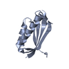 1cqnC  1risS S: Starting model for refinement C: citing same article ( |
|---|---|
| Similar structure data |


-Links
 PDBj
PDBj


- Assembly
| Deposited unit | 
| ||||||||||||
|---|---|---|---|---|---|---|---|---|---|---|---|---|---|
| 1 | 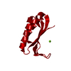
| ||||||||||||
| Unit cell |
| ||||||||||||
| Components on special symmetry positions |
|
-Components
| #1: Protein | Mass: 11830.700 Da / Num. of mol.: 1 / Mutation: YES Source method: isolated from a genetically manipulated source Source: (gene. exp.) Thermus thermophilus (bacteria) / Gene: rpsF, rps6 / Production host: |
|---|---|
| #2: Chemical | ChemComp-MG /   Mass: 24.305 Da / Num. of mol.: 1 / Source method: obtained synthetically / Formula: Mg Mass: 24.305 Da / Num. of mol.: 1 / Source method: obtained synthetically / Formula: Mg |
| #3: Water | ChemComp-HOH /  Mass: 18.015 Da / Num. of mol.: 45 / Source method: isolated from a natural source / Formula: H2O Mass: 18.015 Da / Num. of mol.: 45 / Source method: isolated from a natural source / Formula: H2O |
-Experimental details
-Experiment
| Experiment | Method: X-RAY DIFFRACTION / Number of used crystals: 1 |
|---|
- Sample preparation
| Crystal | Density Matthews: 1.9 Å3/Da / Density % sol: 35 % | ||||||||||||||||||||||||||||||
|---|---|---|---|---|---|---|---|---|---|---|---|---|---|---|---|---|---|---|---|---|---|---|---|---|---|---|---|---|---|---|---|
| Crystal grow | pH: 7 / Details: pH 7.00 | ||||||||||||||||||||||||||||||
| Crystal | *PLUS | ||||||||||||||||||||||||||||||
| Crystal grow | *PLUS Method: unknown / pH: 8.5 | ||||||||||||||||||||||||||||||
| Components of the solutions | *PLUS
|
-Data collection
| Diffraction | Mean temperature: 100 K |
|---|---|
| Diffraction source | Source: SYNCHROTRON / Site: MAX II  / Beamline: I711 / Wavelength: 0.995 / Beamline: I711 / Wavelength: 0.995 |
| Detector | Type: MARRESEARCH / Detector: IMAGE PLATE / Date: Feb 15, 1999 / Details: BENDABLE MIRROR |
| Radiation | Monochromator: SI(111) / Protocol: SINGLE WAVELENGTH / Monochromatic (M) / Laue (L): M / Scattering type: x-ray |
| Radiation wavelength | Wavelength: 0.995 Å / Relative weight: 1 |
| Reflection | Resolution: 2.2→50 Å / Num. obs: 5041 / % possible obs: 99.8 % / Redundancy: 11.6 % / Biso Wilson estimate: 17 Å2 / Rmerge(I) obs: 0.047 / Rsym value: 0.047 / Net I/σ(I): 35 |
| Reflection shell | Resolution: 2.2→2.28 Å / Redundancy: 11.6 % / Rmerge(I) obs: 0.226 / Mean I/σ(I) obs: 11 / Rsym value: 0.226 / % possible all: 100 |
- Processing
| Software |
| ||||||||||||||||||||||||||||||||||||||||||||||||||||||||||||
|---|---|---|---|---|---|---|---|---|---|---|---|---|---|---|---|---|---|---|---|---|---|---|---|---|---|---|---|---|---|---|---|---|---|---|---|---|---|---|---|---|---|---|---|---|---|---|---|---|---|---|---|---|---|---|---|---|---|---|---|---|---|
| Refinement | Method to determine structure: MOLECULAR REPLACEMENT Starting model: 1RIS Resolution: 2.2→35 Å / Rfactor Rfree error: 0.012 / Data cutoff high absF: 2132615.58 / Isotropic thermal model: RESTRAINED / Cross valid method: THROUGHOUT / σ(F): 0
| ||||||||||||||||||||||||||||||||||||||||||||||||||||||||||||
| Solvent computation | Solvent model: FLAT MODEL / Bsol: 65.2756 Å2 / ksol: 0.384563 e/Å3 | ||||||||||||||||||||||||||||||||||||||||||||||||||||||||||||
| Displacement parameters | Biso mean: 34.7 Å2
| ||||||||||||||||||||||||||||||||||||||||||||||||||||||||||||
| Refine analyze |
| ||||||||||||||||||||||||||||||||||||||||||||||||||||||||||||
| Refinement step | Cycle: LAST / Resolution: 2.2→35 Å
| ||||||||||||||||||||||||||||||||||||||||||||||||||||||||||||
| Refine LS restraints |
| ||||||||||||||||||||||||||||||||||||||||||||||||||||||||||||
| LS refinement shell | Resolution: 2.2→2.34 Å / Rfactor Rfree error: 0.034 / Total num. of bins used: 6
| ||||||||||||||||||||||||||||||||||||||||||||||||||||||||||||
| Xplor file |
| ||||||||||||||||||||||||||||||||||||||||||||||||||||||||||||
| Software | *PLUS Name: CNS / Version: 0.5 / Classification: refinement | ||||||||||||||||||||||||||||||||||||||||||||||||||||||||||||
| Refinement | *PLUS σ(F): 0 | ||||||||||||||||||||||||||||||||||||||||||||||||||||||||||||
| Solvent computation | *PLUS | ||||||||||||||||||||||||||||||||||||||||||||||||||||||||||||
| Displacement parameters | *PLUS | ||||||||||||||||||||||||||||||||||||||||||||||||||||||||||||
| Refine LS restraints | *PLUS
|
