 Movie
Movie Controller

[English] 日本語
 Yorodumi
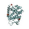
- PDB-6i6s: Circular permutant of ribosomal protein S6, adding 9aa to C termi... -
+ Open data

- Basic information
| Entry | Database: PDB / ID: 6i6s | |||||||||
|---|---|---|---|---|---|---|---|---|---|---|
| Title | Circular permutant of ribosomal protein S6, adding 9aa to C terminal of P68-69, L75A mutant | |||||||||
 Components Components | 30S ribosomal protein S6,30S ribosomal protein S6,30S ribosomal protein S6,30S ribosomal protein S6,30S ribosomal protein S6,30S ribosomal protein S6,30S ribosomal protein S6,30S ribosomal protein S6 | |||||||||
 Keywords Keywords | RIBOSOMAL PROTEIN / Circular permutant / strand swap / local unfolding / cis-proline. / designed protein | |||||||||
| Function / homology |  Function and homology information Function and homology informationsmall ribosomal subunit rRNA binding / structural constituent of ribosome / ribosome / translation / ribonucleoprotein complex / cytoplasm Similarity search - Function | |||||||||
| Biological species |   Thermus thermophilus (bacteria)Thermus thermophilus HB8 (bacteria) Thermus thermophilus (bacteria)Thermus thermophilus HB8 (bacteria) | |||||||||
| Method |  X-RAY DIFFRACTION / SYNCHROTRON / MOLECULAR REPLACEMENT / Resolution: 1.46 Å X-RAY DIFFRACTION / SYNCHROTRON / MOLECULAR REPLACEMENT / Resolution: 1.46 Å | |||||||||
 Authors Authors | Wang, H. / Logan, D.T. / Oliveberg, M. | |||||||||
 Citation Citation | Journal: Proc.Natl.Acad.Sci.USA / Year: 2020 Title: Exposing the distinctive modular behavior of beta-strands and alpha-helices in folded proteins. Authors: Wang, H. / Logan, D.T. / Danielsson, J. / Oliveberg, M. | |||||||||
| History |
|
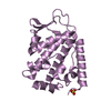
- Structure visualization
| Structure viewer | Molecule: MolmilJmol/JSmol |
|---|
- Downloads & links
-Download
| PDBx/mmCIF format | 6i6s.cif.gz | 133 KB | Display | PDBx/mmCIF format |
|---|---|---|---|---|
| PDB format | pdb6i6s.ent.gz | 105 KB | Display | PDB format |
| PDBx/mmJSON format | 6i6s.json.gz | Tree view | PDBx/mmJSON format | |
| Others |  Other downloads Other downloads |
-Validation report
| Arichive directory | https://data.pdbj.org/pub/pdb/validation_reports/i6/6i6sftp://data.pdbj.org/pub/pdb/validation_reports/i6/6i6s | HTTPS FTP |
|---|
-Related structure data
| Related structure data |  6i69C  6i6eC  6i6iC  6i6oC 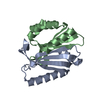 6i6uC  6i6wC  6i6yC  1risS S: Starting model for refinement C: citing same article ( |
|---|---|
| Similar structure data |


-Links
 PDBj
PDBj


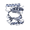
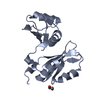
- Assembly
| Deposited unit | 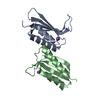
| ||||||||
|---|---|---|---|---|---|---|---|---|---|
| 1 | 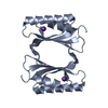
| ||||||||
| 2 | 
| ||||||||
| Unit cell |
|
-Components
| #1: Protein | Mass: 12287.949 Da / Num. of mol.: 2 Source method: isolated from a genetically manipulated source Source: (gene. exp.) Thermus thermophilus (strain HB8 / ATCC 27634 / DSM 579) (bacteria), (gene. exp.) Thermus thermophilus HB8 (bacteria)Strain: HB8 / ATCC 27634 / DSM 579 / Gene: rpsF, TTHA0245 / Production host: #2: Chemical | ChemComp-K / |   Mass: 39.098 Da / Num. of mol.: 1 / Source method: isolated from a natural source / Formula: K Mass: 39.098 Da / Num. of mol.: 1 / Source method: isolated from a natural source / Formula: K#3: Chemical | ChemComp-NA / |   Mass: 22.990 Da / Num. of mol.: 1 / Source method: obtained synthetically / Formula: Na Mass: 22.990 Da / Num. of mol.: 1 / Source method: obtained synthetically / Formula: Na#4: Water | ChemComp-HOH / |  Mass: 18.015 Da / Num. of mol.: 102 / Source method: isolated from a natural source / Formula: H2O Mass: 18.015 Da / Num. of mol.: 102 / Source method: isolated from a natural source / Formula: H2OHas ligand of interest | N | |
|---|
-Experimental details
-Experiment
| Experiment | Method: X-RAY DIFFRACTION / Number of used crystals: 1 |
|---|
- Sample preparation
| Crystal | Density Matthews: 1.91 Å3/Da / Density % sol: 35.71 % |
|---|---|
| Crystal grow | Temperature: 293 K / Method: vapor diffusion, sitting drop Details: 0.2 M Potassium sodium tartrate tetrahydrate 0.1 M Bis-Tris propane pH 7.5 20% w/v PEG 3350 |
-Data collection
| Diffraction | Mean temperature: 100 K / Serial crystal experiment: N | ||||||||||||||||||||||||
|---|---|---|---|---|---|---|---|---|---|---|---|---|---|---|---|---|---|---|---|---|---|---|---|---|---|
| Diffraction source | Source: SYNCHROTRON / Site: MAX IV  / Beamline: BioMAX / Wavelength: 0.97999 Å / Beamline: BioMAX / Wavelength: 0.97999 Å | ||||||||||||||||||||||||
| Detector | Type: DECTRIS EIGER X 16M / Detector: PIXEL / Date: Jan 31, 2018 | ||||||||||||||||||||||||
| Radiation | Protocol: SINGLE WAVELENGTH / Monochromatic (M) / Laue (L): M / Scattering type: x-ray | ||||||||||||||||||||||||
| Radiation wavelength | Wavelength: 0.97999 Å / Relative weight: 1 | ||||||||||||||||||||||||
| Reflection | Resolution: 1.46→133.06 Å / Num. obs: 30436 / % possible obs: 100 % / Redundancy: 12.7 % / CC1/2: 0.998 / Rmerge(I) obs: 0.1 / Rpim(I) all: 0.029 / Rrim(I) all: 0.104 / Net I/σ(I): 11.6 / Num. measured all: 387834 / Scaling rejects: 17 | ||||||||||||||||||||||||
| Reflection shell | Diffraction-ID: 1
|
- Processing
| Software |
| ||||||||||||||||||||||||||||||||||||||||||||||||||||||||||||||||||||||||
|---|---|---|---|---|---|---|---|---|---|---|---|---|---|---|---|---|---|---|---|---|---|---|---|---|---|---|---|---|---|---|---|---|---|---|---|---|---|---|---|---|---|---|---|---|---|---|---|---|---|---|---|---|---|---|---|---|---|---|---|---|---|---|---|---|---|---|---|---|---|---|---|---|---|
| Refinement | Method to determine structure: MOLECULAR REPLACEMENT Starting model: 1Ris Resolution: 1.46→35.84 Å / SU ML: 0.14 / Cross valid method: THROUGHOUT / σ(F): 1.35 / Phase error: 19.66 / Stereochemistry target values: ML
| ||||||||||||||||||||||||||||||||||||||||||||||||||||||||||||||||||||||||
| Solvent computation | Shrinkage radii: 0.9 Å / VDW probe radii: 1.11 Å / Solvent model: FLAT BULK SOLVENT MODEL | ||||||||||||||||||||||||||||||||||||||||||||||||||||||||||||||||||||||||
| Displacement parameters | Biso max: 104.28 Å2 / Biso mean: 34.0085 Å2 / Biso min: 15.8 Å2 | ||||||||||||||||||||||||||||||||||||||||||||||||||||||||||||||||||||||||
| Refinement step | Cycle: final / Resolution: 1.46→35.84 Å
| ||||||||||||||||||||||||||||||||||||||||||||||||||||||||||||||||||||||||
| LS refinement shell | Refine-ID: X-RAY DIFFRACTION / Rfactor Rfree error: 0 / Total num. of bins used: 11 / % reflection obs: 100 %
|
