 Movie
Movie Controller


+ Open data

- Basic information
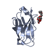
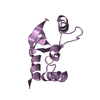
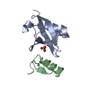
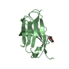
| Entry | Database: PDB / ID: 2bxj | ||||||
|---|---|---|---|---|---|---|---|
| Title | Double Mutant of the Ribosomal Protein S6 | ||||||
 Components Components | 30S RIBOSOMAL PROTEIN S6 | ||||||
 Keywords Keywords | RIBOSOMAL PROTEIN / S6 DOUBLE MUTANT / RNA-BINDING | ||||||
| Function / homology |  Function and homology information Function and homology informationsmall ribosomal subunit rRNA binding / structural constituent of ribosome / ribosome / translation / ribonucleoprotein complex / cytoplasm Similarity search - Function | ||||||
| Biological species |   THERMUS THERMOPHILUS (bacteria) THERMUS THERMOPHILUS (bacteria) | ||||||
| Method |  X-RAY DIFFRACTION / MOLECULAR REPLACEMENT / Resolution: 2.4 Å X-RAY DIFFRACTION / MOLECULAR REPLACEMENT / Resolution: 2.4 Å | ||||||
 Authors Authors | Otzen, D.E. | ||||||
 Citation Citation | Journal: Protein Eng.Des.Sel. / Year: 2005 Title: Antagonism, Non-Native Interactions and Non-Two-State Folding in S6 Revealed by Double-Mutant Cycle Analysis. Authors: Otzen, D.E. #1: Journal: Biochemistry / Year: 1999Title: Structural Changes in the Transition State of Protein Folding: Alternative Interpretations of Curved Chevron Plots Authors: Otzen, D.E. / Kristensen, O. / Proctor, M. / Oliveberg, M. #2: Journal: Biochemistry / Year: 1997 Title: High-Energy Channelling in Protein Folding Authors: Silow, M. / Oliveberg, M. #3: Journal: Embo J. / Year: 1994Title: Crystal Structure of the Ribosomal Protein S6 from Thermus Thermophilus Authors: Lindahl, M. / Svensson, L.A. / Liljas, A. / Sedelnikova, S.E. / Eliseikina, I.A. / Fomenkova, N.P. / Nevskaya, N. / Nikonov, S.V. / Garber, M.B. / Muranova, T.A. / Rykonova, A.I. / Amons, R. | ||||||
| History |
| ||||||
| Remark 650 | HELIX DETERMINATION METHOD: AUTHOR PROVIDED. |
- Structure visualization
| Structure viewer | Molecule: MolmilJmol/JSmol |
|---|
- Downloads & links
-Download
| PDBx/mmCIF format | 2bxj.cif.gz | 53.3 KB | Display | PDBx/mmCIF format |
|---|---|---|---|---|
| PDB format | pdb2bxj.ent.gz | 39.1 KB | Display | PDB format |
| PDBx/mmJSON format | 2bxj.json.gz | Tree view | PDBx/mmJSON format | |
| Others |  Other downloads Other downloads |
-Validation report
| Arichive directory | https://data.pdbj.org/pub/pdb/validation_reports/bx/2bxjftp://data.pdbj.org/pub/pdb/validation_reports/bx/2bxj | HTTPS FTP |
|---|
-Related structure data
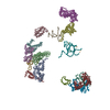
| Related structure data | 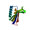 2bvzC  1louS S: Starting model for refinement C: citing same article ( |
|---|---|
| Similar structure data |


-Links
 PDBj
PDBj


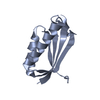
- Assembly
| Deposited unit | 
| ||||||||
|---|---|---|---|---|---|---|---|---|---|
| 1 | 
| ||||||||
| 2 | 
| ||||||||
| Unit cell |
|
-Components
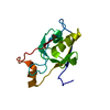
| #1: Protein | Mass: 11904.593 Da / Num. of mol.: 2 / Mutation: YES Source method: isolated from a genetically manipulated source Source: (gene. exp.) THERMUS THERMOPHILUS (bacteria) / Production host: #2: Water | ChemComp-HOH / |  Mass: 18.015 Da / Num. of mol.: 70 / Source method: isolated from a natural source / Formula: H2O Mass: 18.015 Da / Num. of mol.: 70 / Source method: isolated from a natural source / Formula: H2OCompound details | ENGINEERED | Sequence details | DOUBLE MUTANT OF P23370 (L30A, L75A) | |
|---|
-Experimental details
-Experiment
| Experiment | Method: X-RAY DIFFRACTION / Number of used crystals: 1 |
|---|
- Sample preparation
| Crystal | Density Matthews: 2.06 Å3/Da / Density % sol: 40.24 % |
|---|---|
| Crystal grow | Details: 0.2M NA-CITRATE, 0.1M TRIS-CL PH 8.5, 25% (V/V) PEG400 |
-Data collection
| Diffraction | Mean temperature: 100 K |
|---|---|
| Diffraction source | Source: ROTATING ANODE / Type: RIGAKU RU200 / Wavelength: 1.5418 |
| Detector | Type: MARRESEARCH / Detector: IMAGE PLATE |
| Radiation | Monochromator: GRAPHITE / Protocol: SINGLE WAVELENGTH / Monochromatic (M) / Laue (L): M / Scattering type: x-ray |
| Radiation wavelength | Wavelength: 1.5418 Å / Relative weight: 1 |
| Reflection | Resolution: 2.4→23.05 Å / Num. obs: 12515 / % possible obs: 97 % / Observed criterion σ(I): 3 / Redundancy: 18 % / Biso Wilson estimate: 20 Å2 / Rmerge(I) obs: 0.12 / Net I/σ(I): 23.56 |
| Reflection shell | Resolution: 2.4→2.55 Å / Redundancy: 12 % / Rmerge(I) obs: 0.43 / Mean I/σ(I) obs: 6.9 / % possible all: 95.2 |
- Processing
| Software |
| ||||||||||||||||||||||||||||||||||||||||||||||||||||||||||||||||||||||||||||||||
|---|---|---|---|---|---|---|---|---|---|---|---|---|---|---|---|---|---|---|---|---|---|---|---|---|---|---|---|---|---|---|---|---|---|---|---|---|---|---|---|---|---|---|---|---|---|---|---|---|---|---|---|---|---|---|---|---|---|---|---|---|---|---|---|---|---|---|---|---|---|---|---|---|---|---|---|---|---|---|---|---|---|
| Refinement | Method to determine structure: MOLECULAR REPLACEMENT Starting model: PDB ENTRY 1LOU Resolution: 2.4→23.05 Å / Rfactor Rfree error: 0.009 / Data cutoff high absF: 261301.8 / Isotropic thermal model: RESTRAINED / Cross valid method: THROUGHOUT / σ(F): 0
| ||||||||||||||||||||||||||||||||||||||||||||||||||||||||||||||||||||||||||||||||
| Solvent computation | Solvent model: FLAT MODEL / Bsol: 40.8353 Å2 / ksol: 0.321317 e/Å3 | ||||||||||||||||||||||||||||||||||||||||||||||||||||||||||||||||||||||||||||||||
| Displacement parameters | Biso mean: 27.3 Å2
| ||||||||||||||||||||||||||||||||||||||||||||||||||||||||||||||||||||||||||||||||
| Refine analyze |
| ||||||||||||||||||||||||||||||||||||||||||||||||||||||||||||||||||||||||||||||||
| Refinement step | Cycle: LAST / Resolution: 2.4→23.05 Å
| ||||||||||||||||||||||||||||||||||||||||||||||||||||||||||||||||||||||||||||||||
| Refine LS restraints |
| ||||||||||||||||||||||||||||||||||||||||||||||||||||||||||||||||||||||||||||||||
| LS refinement shell | Resolution: 2.4→2.55 Å / Rfactor Rfree error: 0.031 / Total num. of bins used: 6
| ||||||||||||||||||||||||||||||||||||||||||||||||||||||||||||||||||||||||||||||||
| Xplor file |
|
