 Movie
Movie Controller


+ Open data

- Basic information
| Entry | Database: PDB / ID: 3r15 | ||||||
|---|---|---|---|---|---|---|---|
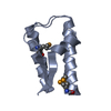
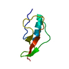
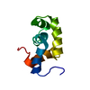
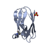
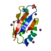
| Title | Structure Treponema Denticola Factor H Binding Protein | ||||||
 Components Components | Factor H binding protein | ||||||
 Keywords Keywords | PROTEIN BINDING / Factor H binding protein / Factor H | ||||||
| Function / homology |  Function and homology information Function and homology informationSingle alpha-helices involved in coiled-coils or other helix-helix interfaces - #2300 / : / : / Factor H binding protein B / Single alpha-helices involved in coiled-coils or other helix-helix interfaces / Helix non-globular / Special / Prokaryotic membrane lipoprotein lipid attachment site profile. Similarity search - Domain/homology | ||||||
| Biological species |  Treponema denticola (bacteria) Treponema denticola (bacteria) | ||||||
| Method |  X-RAY DIFFRACTION / SYNCHROTRON / MOLECULAR REPLACEMENT / Resolution: 1.701 Å X-RAY DIFFRACTION / SYNCHROTRON / MOLECULAR REPLACEMENT / Resolution: 1.701 Å | ||||||
 Authors Authors | Miller, D.P. / McDowell, J.V. / Burgner, J. / Heroux, A. / Bell, J.K. / Marconi, R.T. | ||||||
 Citation Citation | Journal: To be Published Title: Structure Treponema Denticola Factor H Binding Protein Authors: Miller, D.P. / McDowell, J.V. / Burgner, J. / Heroux, A. / Bell, J.K. / Marconi, R.T. #1: Journal: To be PublishedTitle: Crystallization of the Factor H binding protein, FhbB, of the periopathogen Treponema denticola Authors: Miller, D.P. / McDowell, J.V. / Bell, J.K. / Marconi, R.T. #2: Journal: Infect.Immun. / Year: 2009Title: Analysis of a unique interaction between the complement regulatory protein factor H and the periodontal pathogen Treponema denticola Authors: McDowell, J.V. / Huang, B. / Fenno, J.C. / Marconi, R.T. | ||||||
| History |
|
- Structure visualization
| Structure viewer | Molecule: MolmilJmol/JSmol |
|---|
- Downloads & links
-Download
| PDBx/mmCIF format | 3r15.cif.gz | 46.1 KB | Display | PDBx/mmCIF format |
|---|---|---|---|---|
| PDB format | pdb3r15.ent.gz | 32 KB | Display | PDB format |
| PDBx/mmJSON format | 3r15.json.gz | Tree view | PDBx/mmJSON format | |
| Others |  Other downloads Other downloads |
-Validation report
| Arichive directory | https://data.pdbj.org/pub/pdb/validation_reports/r1/3r15ftp://data.pdbj.org/pub/pdb/validation_reports/r1/3r15 | HTTPS FTP |
|---|
-Related structure data
| Related structure data |  3qz0S S: Starting model for refinement |
|---|---|
| Similar structure data |


-Links
 PDBj
PDBj


- Assembly
| Deposited unit | 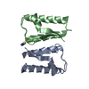
| ||||||||||||
|---|---|---|---|---|---|---|---|---|---|---|---|---|---|
| 1 | 
| ||||||||||||
| 2 | 
| ||||||||||||
| Unit cell |
| ||||||||||||
| Components on special symmetry positions |
|
-Components
| #1: Protein | Mass: 10560.178 Da / Num. of mol.: 2 / Fragment: residues 24-102 Source method: isolated from a genetically manipulated source Source: (gene. exp.) Treponema denticola (bacteria) / Strain: ATCC 35405 / Gene: fhbB, TDE_0108 / Production host: #2: Chemical |   Mass: 58.082 Da / Num. of mol.: 2 / Source method: obtained synthetically / Formula: CNS Mass: 58.082 Da / Num. of mol.: 2 / Source method: obtained synthetically / Formula: CNS#3: Water | ChemComp-HOH / |  Mass: 18.015 Da / Num. of mol.: 117 / Source method: isolated from a natural source / Formula: H2O Mass: 18.015 Da / Num. of mol.: 117 / Source method: isolated from a natural source / Formula: H2O |
|---|
-Experimental details
-Experiment
| Experiment | Method: X-RAY DIFFRACTION / Number of used crystals: 1 |
|---|
- Sample preparation
| Crystal | Density Matthews: 2.21 Å3/Da / Density % sol: 44.28 % |
|---|---|
| Crystal grow | Temperature: 291 K / Method: vapor diffusion, hanging drop / pH: 7.5 Details: 0.1 M HEPES, 1.2 M sodium citrate tribasic dihydrate, 0.2 M sodium thiocyanate, 2.5% glycerol, pH 7.5, VAPOR DIFFUSION, HANGING DROP, temperature 291K |
-Data collection
| Diffraction | Mean temperature: 170 K |
|---|---|
| Diffraction source | Source: SYNCHROTRON / Site: NSLS  / Beamline: X25 / Wavelength: 1.5 Å / Beamline: X25 / Wavelength: 1.5 Å |
| Detector | Type: ADSC QUANTUM 315 / Detector: CCD / Date: Mar 18, 2010 |
| Radiation | Monochromator: Double silicon(111) crystal monochromator with cryogenically-cooled first crystal and sagittally-bent second crystal horizontally-focusing at 3.3:1 demagnification Protocol: SINGLE WAVELENGTH / Monochromatic (M) / Laue (L): M / Scattering type: x-ray |
| Radiation wavelength | Wavelength: 1.5 Å / Relative weight: 1 |
| Reflection | Resolution: 1.7→40 Å / Num. all: 39634 / Num. obs: 39492 / % possible obs: 99.6 % / Observed criterion σ(F): 2 / Observed criterion σ(I): 2 / Redundancy: 5.4 % |
- Processing
| Software |
| |||||||||||||||||||||||||||||||||||||||||||||||||||||||||||||||||||||||||||||
|---|---|---|---|---|---|---|---|---|---|---|---|---|---|---|---|---|---|---|---|---|---|---|---|---|---|---|---|---|---|---|---|---|---|---|---|---|---|---|---|---|---|---|---|---|---|---|---|---|---|---|---|---|---|---|---|---|---|---|---|---|---|---|---|---|---|---|---|---|---|---|---|---|---|---|---|---|---|---|
| Refinement | Method to determine structure: MOLECULAR REPLACEMENT Starting model: 3QZ0 Resolution: 1.701→36.079 Å / SU ML: 0.19 / σ(F): 0.15 / Stereochemistry target values: ML
| |||||||||||||||||||||||||||||||||||||||||||||||||||||||||||||||||||||||||||||
| Solvent computation | Shrinkage radii: 0.9 Å / VDW probe radii: 1.11 Å / Solvent model: FLAT BULK SOLVENT MODEL / Bsol: 43.074 Å2 / ksol: 0.383 e/Å3 | |||||||||||||||||||||||||||||||||||||||||||||||||||||||||||||||||||||||||||||
| Displacement parameters |
| |||||||||||||||||||||||||||||||||||||||||||||||||||||||||||||||||||||||||||||
| Refinement step | Cycle: LAST / Resolution: 1.701→36.079 Å
| |||||||||||||||||||||||||||||||||||||||||||||||||||||||||||||||||||||||||||||
| Refine LS restraints |
| |||||||||||||||||||||||||||||||||||||||||||||||||||||||||||||||||||||||||||||
| LS refinement shell |
|
