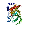
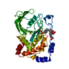
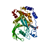
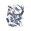
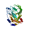
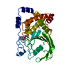
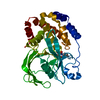
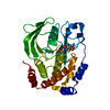
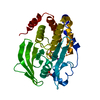
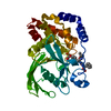
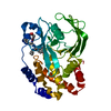
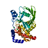
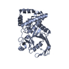
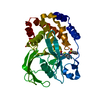
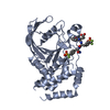
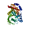
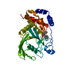
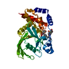
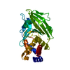
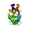
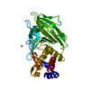
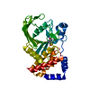
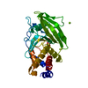
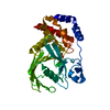
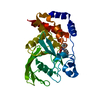
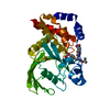
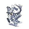
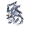
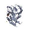
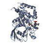
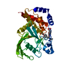
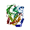
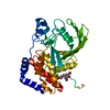
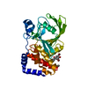
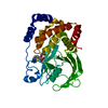
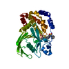
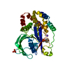
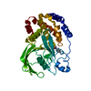
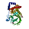
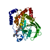
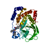
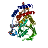
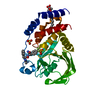
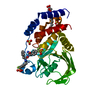
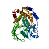
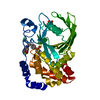
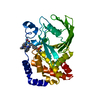
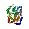
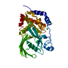
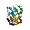
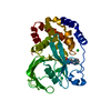
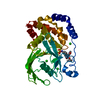
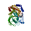
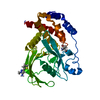
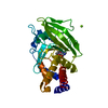
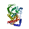
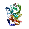
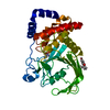
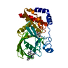
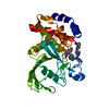
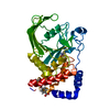
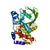
Entry Database : PDB / ID : 3zv2Title Human protein-tyrosine phosphatase 1b C215A, S216A mutant Tyrosine-protein phosphatase non-receptor type 1 Keywords / Function / homology Function Domain/homology Component
/ / / / / / / / / / / / / / / / / / / / / / / / / / / / / / / / / / / / / / / / / / / / / / / / / / / / / / / / / / / / / / / / / / / / / / / / / / / / / / / / / / / / / / / / / / / / / / / / / / / / / / / / / / / / / / Biological species Homo sapiens (human)Method / / Resolution : 2.8 Å Authors Barford, D. / Salmeen, A. / Tonks, N.K. Journal : Cell / Year : 2011Title : Conformation-sensing antibodies stabilize the oxidized form of PTP1B and inhibit its phosphatase activity.Authors : Haque, A. / Andersen, J.N. / Salmeen, A. / Barford, D. / Tonks, N.K. History Deposition Jul 22, 2011 Deposition site / Processing site Revision 1.0 Aug 31, 2011 Provider / Type Revision 1.1 Oct 12, 2011 Group Revision 1.2 Jul 25, 2018 Group Data collection / Database references ... Data collection / Database references / Source and taxonomy / Structure summary Category citation / entity ... citation / entity / entity_name_com / entity_src_gen / entity_src_nat / struct_ref Item _citation.country / _citation.journal_abbrev ... _citation.country / _citation.journal_abbrev / _citation.journal_id_ASTM / _citation.journal_id_CSD / _citation.journal_id_ISSN / _citation.page_last / _citation.pdbx_database_id_DOI / _citation.title / _entity.pdbx_description / _entity.pdbx_mutation / _entity.src_method / _entity_name_com.name / _struct_ref.pdbx_align_begin / _struct_ref.pdbx_seq_one_letter_code Revision 1.3 Dec 20, 2023 Group Data collection / Database references ... Data collection / Database references / Other / Refinement description Category chem_comp_atom / chem_comp_bond ... chem_comp_atom / chem_comp_bond / database_2 / pdbx_database_status / pdbx_initial_refinement_model Item / _database_2.pdbx_database_accession / _pdbx_database_status.status_code_sf
Show all Show less
 Movie
Movie Controller


 Open data
 Basic information
 Components
 Keywords
 Function and homology information
 Homo sapiens (human)
 X-RAY DIFFRACTION /
  Authors
 Citation
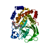
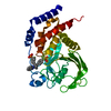
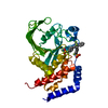
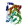
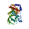
 Structure visualization
 Downloads & links
 Other downloads


 PDBj
PDBj


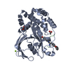
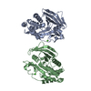
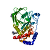
 Assembly

 Sample preparation
 Processing