 Movie
Movie Controller


+ Open data

- Basic information
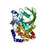
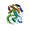
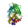
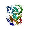
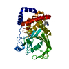
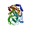
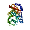
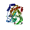
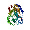
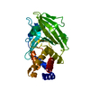
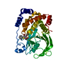
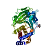
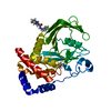
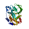
| Entry | Database: PDB / ID: 1bzh | |||||||||
|---|---|---|---|---|---|---|---|---|---|---|
| Title | Cyclic peptide inhibitor of human PTP1B | |||||||||
 Components Components |
| |||||||||
 Keywords Keywords | HYDROLASE/HYDROLASE INHIBITOR / PHOSPHORYLATION-INHIBITOR COMPLEX / TYROSINE PHOSPHATASE / HYDROLASE-HYDROLASE INHIBITOR complex | |||||||||
| Function / homology |  Function and homology information Function and homology informationPTK6 Down-Regulation / regulation of hepatocyte growth factor receptor signaling pathway / positive regulation of receptor catabolic process / insulin receptor recycling / negative regulation of vascular endothelial growth factor receptor signaling pathway / regulation of intracellular protein transport / negative regulation of MAP kinase activity / mitochondrial crista / IRE1-mediated unfolded protein response / platelet-derived growth factor receptor-beta signaling pathway ...PTK6 Down-Regulation / regulation of hepatocyte growth factor receptor signaling pathway / positive regulation of receptor catabolic process / insulin receptor recycling / negative regulation of vascular endothelial growth factor receptor signaling pathway / regulation of intracellular protein transport / negative regulation of MAP kinase activity / mitochondrial crista / IRE1-mediated unfolded protein response / platelet-derived growth factor receptor-beta signaling pathway / sorting endosome / positive regulation of IRE1-mediated unfolded protein response / negative regulation of PERK-mediated unfolded protein response / regulation of type I interferon-mediated signaling pathway / cytoplasmic side of endoplasmic reticulum membrane / negative regulation of vascular associated smooth muscle cell migration / vascular endothelial cell response to oscillatory fluid shear stress / peptidyl-tyrosine dephosphorylation / positive regulation of systemic arterial blood pressure / non-membrane spanning protein tyrosine phosphatase activity / Regulation of IFNA/IFNB signaling / regulation of endocytosis / cellular response to angiotensin / regulation of proteolysis / growth hormone receptor signaling pathway via JAK-STAT / negative regulation of cell-substrate adhesion / cellular response to unfolded protein / regulation of postsynapse assembly / positive regulation of endothelial cell apoptotic process / regulation of signal transduction / negative regulation of signal transduction / Regulation of IFNG signaling / Growth hormone receptor signaling / negative regulation of endoplasmic reticulum stress-induced intrinsic apoptotic signaling pathway / positive regulation of heart rate / ephrin receptor binding / cellular response to platelet-derived growth factor stimulus / Insulin receptor recycling / MECP2 regulates neuronal receptors and channels / Integrin signaling / endoplasmic reticulum unfolded protein response / phosphoprotein phosphatase activity / protein-tyrosine-phosphatase / cellular response to fibroblast growth factor stimulus / cellular response to nitric oxide / negative regulation of insulin receptor signaling pathway / positive regulation of cardiac muscle cell apoptotic process / protein tyrosine phosphatase activity / protein phosphatase 2A binding / Turbulent (oscillatory, disturbed) flow shear stress activates signaling by PIEZO1 and integrins in endothelial cells / endosome lumen / negative regulation of phosphatidylinositol 3-kinase/protein kinase B signal transduction / insulin receptor binding / cellular response to nerve growth factor stimulus / negative regulation of ERK1 and ERK2 cascade / response to nutrient levels / Negative regulation of MET activity / receptor tyrosine kinase binding / positive regulation of JNK cascade / insulin receptor signaling pathway / negative regulation of neuron projection development / actin cytoskeleton organization / cellular response to hypoxia / early endosome / postsynapse / cadherin binding / mitochondrial matrix / negative regulation of cell population proliferation / protein kinase binding / glutamatergic synapse / enzyme binding / endoplasmic reticulum / protein-containing complex / RNA binding / zinc ion binding / cytosol / cytoplasm Similarity search - Function | |||||||||
| Biological species |  Homo sapiens (human) Homo sapiens (human) | |||||||||
| Method |  X-RAY DIFFRACTION / SYNCHROTRON / MOLECULAR REPLACEMENT / Resolution: 2.1 Å X-RAY DIFFRACTION / SYNCHROTRON / MOLECULAR REPLACEMENT / Resolution: 2.1 Å | |||||||||
 Authors Authors | Groves, M.R. / Yao, Z.J. / Burke Jr., T.R. / Barford, D. | |||||||||
 Citation Citation | Journal: Biochemistry / Year: 1998 Title: Structural basis for inhibition of the protein tyrosine phosphatase 1B by phosphotyrosine peptide mimetics. Authors: Groves, M.R. / Yao, Z.J. / Roller, P.P. / Burke Jr., T.R. / Barford, D. #1: Journal: Science / Year: 1994Title: Crystal Structure of Human Protein Tyrosine Phosphatase 1B Authors: Barford, D. / Flint, A.J. / Tonks, N.K. | |||||||||
| History |
|
- Structure visualization
| Structure viewer | Molecule: MolmilJmol/JSmol |
|---|
- Downloads & links
-Download
| PDBx/mmCIF format | 1bzh.cif.gz | 77.5 KB | Display | PDBx/mmCIF format |
|---|---|---|---|---|
| PDB format | pdb1bzh.ent.gz | 56.4 KB | Display | PDB format |
| PDBx/mmJSON format | 1bzh.json.gz | Tree view | PDBx/mmJSON format | |
| Others |  Other downloads Other downloads |
-Validation report
| Arichive directory | https://data.pdbj.org/pub/pdb/validation_reports/bz/1bzhftp://data.pdbj.org/pub/pdb/validation_reports/bz/1bzh | HTTPS FTP |
|---|
-Related structure data
| Related structure data | 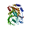 1bzcC 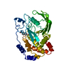 1bzjC 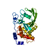 2hnpS S: Starting model for refinement C: citing same article ( |
|---|---|
| Similar structure data |


-Links
 PDBj
PDBj


- Assembly
| Deposited unit | 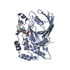
| ||||||||
|---|---|---|---|---|---|---|---|---|---|
| 1 |
| ||||||||
| Unit cell |
|
-Components
| #1: Protein | Mass: 34720.566 Da / Num. of mol.: 1 / Fragment: CATALYTIC DOMAIN Source method: isolated from a genetically manipulated source Source: (gene. exp.) Homo sapiens (human) / Production host:  |
|---|---|
| #2: Protein/peptide | Mass: 1004.944 Da / Num. of mol.: 1 Source method: isolated from a genetically manipulated source Source: (gene. exp.) Homo sapiens (human) / Production host: |
| #3: Water | ChemComp-HOH /  Mass: 18.015 Da / Num. of mol.: 88 / Source method: isolated from a natural source / Formula: H2O Mass: 18.015 Da / Num. of mol.: 88 / Source method: isolated from a natural source / Formula: H2O |
-Experimental details
-Experiment
| Experiment | Method: X-RAY DIFFRACTION / Number of used crystals: 1 |
|---|
- Sample preparation
| Crystal | Density Matthews: 2.72 Å3/Da / Density % sol: 54.47 % | ||||||||||||||||||||||||||||||||||||
|---|---|---|---|---|---|---|---|---|---|---|---|---|---|---|---|---|---|---|---|---|---|---|---|---|---|---|---|---|---|---|---|---|---|---|---|---|---|
| Crystal grow | pH: 8 / Details: pH 8.0 | ||||||||||||||||||||||||||||||||||||
| Crystal | *PLUS | ||||||||||||||||||||||||||||||||||||
| Crystal grow | *PLUS Temperature: 4 ℃ / Method: vapor diffusion, hanging drop | ||||||||||||||||||||||||||||||||||||
| Components of the solutions | *PLUS
|
-Data collection
| Diffraction | Mean temperature: 100 K |
|---|---|
| Diffraction source | Source: SYNCHROTRON / Site: ELETTRA  / Beamline: 5.2R / Wavelength: 1 / Beamline: 5.2R / Wavelength: 1 |
| Detector | Type: MAR scanner 345 mm plate / Detector: IMAGE PLATE |
| Radiation | Monochromator: GRAPHITE / Protocol: SINGLE WAVELENGTH / Monochromatic (M) / Laue (L): M / Scattering type: x-ray |
| Radiation wavelength | Wavelength: 1 Å / Relative weight: 1 |
| Reflection | Resolution: 2.1→8 Å / Num. obs: 15934 / % possible obs: 78.1 % / Redundancy: 2.9 % / Rmerge(I) obs: 0.029 / Rsym value: 0.029 / Net I/σ(I): 13.7 |
| Reflection shell | Resolution: 2.1→2.21 Å / Rmerge(I) obs: 0.074 / Mean I/σ(I) obs: 8.8 / Rsym value: 0.074 / % possible all: 55.7 |
| Reflection | *PLUS Num. measured all: 46108 |
| Reflection shell | *PLUS % possible obs: 55.7 % |
- Processing
| Software |
| ||||||||||||||||||||||||||||||||||||||||||||||||||||||||||||||||||||||||||||||||||||
|---|---|---|---|---|---|---|---|---|---|---|---|---|---|---|---|---|---|---|---|---|---|---|---|---|---|---|---|---|---|---|---|---|---|---|---|---|---|---|---|---|---|---|---|---|---|---|---|---|---|---|---|---|---|---|---|---|---|---|---|---|---|---|---|---|---|---|---|---|---|---|---|---|---|---|---|---|---|---|---|---|---|---|---|---|---|
| Refinement | Method to determine structure: MOLECULAR REPLACEMENT Starting model: PDB ENTRY 2HNP Resolution: 2.1→8 Å / Cross valid method: THROUGHOUT / σ(F): 0
| ||||||||||||||||||||||||||||||||||||||||||||||||||||||||||||||||||||||||||||||||||||
| Displacement parameters | Biso mean: 65.49 Å2 | ||||||||||||||||||||||||||||||||||||||||||||||||||||||||||||||||||||||||||||||||||||
| Refinement step | Cycle: LAST / Resolution: 2.1→8 Å
| ||||||||||||||||||||||||||||||||||||||||||||||||||||||||||||||||||||||||||||||||||||
| Refine LS restraints |
| ||||||||||||||||||||||||||||||||||||||||||||||||||||||||||||||||||||||||||||||||||||
| Software | *PLUS Name: REFMAC / Classification: refinement | ||||||||||||||||||||||||||||||||||||||||||||||||||||||||||||||||||||||||||||||||||||
| Refinement | *PLUS Highest resolution: 2.1 Å / σ(F): 0 / % reflection Rfree: 6.67 % / Rfactor obs: 0.211 | ||||||||||||||||||||||||||||||||||||||||||||||||||||||||||||||||||||||||||||||||||||
| Solvent computation | *PLUS | ||||||||||||||||||||||||||||||||||||||||||||||||||||||||||||||||||||||||||||||||||||
| Displacement parameters | *PLUS | ||||||||||||||||||||||||||||||||||||||||||||||||||||||||||||||||||||||||||||||||||||
| Refine LS restraints | *PLUS Type: p_plane_restr / Dev ideal target: 0.03 | ||||||||||||||||||||||||||||||||||||||||||||||||||||||||||||||||||||||||||||||||||||
| LS refinement shell | *PLUS Highest resolution: 2.1 Å / Lowest resolution: 2.21 Å / Rfactor Rfree: 0.336 / Rfactor obs: 0.31 |
