 Movie
Movie Controller

[English] 日本語
 Yorodumi
- PDB-1een: CRYSTAL STRUCTURE OF PROTEIN TYROSINE PHOSPHATASE 1B COMPLEXED WI... -
+ Open data

- Basic information
| Entry | Database: PDB / ID: 1een | ||||||
|---|---|---|---|---|---|---|---|
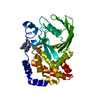
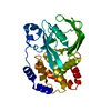
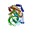
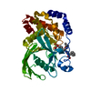
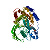
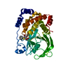
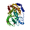
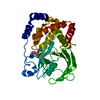
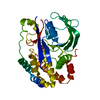
| Title | CRYSTAL STRUCTURE OF PROTEIN TYROSINE PHOSPHATASE 1B COMPLEXED WITH ACETYL-D-A-D-BPA-PTYR-L-I-P-Q-Q-G | ||||||
 Components Components |
| ||||||
 Keywords Keywords | HYDROLASE / ACETYLATION / PHOSPHORYLATION / INHIBITION | ||||||
| Function / homology |  Function and homology information Function and homology informationPTK6 Down-Regulation / regulation of hepatocyte growth factor receptor signaling pathway / positive regulation of receptor catabolic process / insulin receptor recycling / negative regulation of vascular endothelial growth factor receptor signaling pathway / negative regulation of MAP kinase activity / regulation of intracellular protein transport / mitochondrial crista / IRE1-mediated unfolded protein response / sorting endosome ...PTK6 Down-Regulation / regulation of hepatocyte growth factor receptor signaling pathway / positive regulation of receptor catabolic process / insulin receptor recycling / negative regulation of vascular endothelial growth factor receptor signaling pathway / negative regulation of MAP kinase activity / regulation of intracellular protein transport / mitochondrial crista / IRE1-mediated unfolded protein response / sorting endosome / platelet-derived growth factor receptor-beta signaling pathway / positive regulation of IRE1-mediated unfolded protein response / cytoplasmic side of endoplasmic reticulum membrane / negative regulation of PERK-mediated unfolded protein response / regulation of type I interferon-mediated signaling pathway / negative regulation of vascular associated smooth muscle cell migration / vascular endothelial cell response to oscillatory fluid shear stress / peptidyl-tyrosine dephosphorylation / positive regulation of systemic arterial blood pressure / non-membrane spanning protein tyrosine phosphatase activity / regulation of endocytosis / Regulation of IFNA/IFNB signaling / cellular response to angiotensin / regulation of proteolysis / growth hormone receptor signaling pathway via JAK-STAT / negative regulation of cell-substrate adhesion / cellular response to unfolded protein / regulation of postsynapse assembly / positive regulation of endothelial cell apoptotic process / regulation of signal transduction / Regulation of IFNG signaling / negative regulation of signal transduction / Growth hormone receptor signaling / negative regulation of endoplasmic reticulum stress-induced intrinsic apoptotic signaling pathway / positive regulation of heart rate / ephrin receptor binding / Insulin receptor recycling / MECP2 regulates neuronal receptors and channels / cellular response to platelet-derived growth factor stimulus / Integrin signaling / endoplasmic reticulum unfolded protein response / phosphoprotein phosphatase activity / protein-tyrosine-phosphatase / cellular response to fibroblast growth factor stimulus / cellular response to nitric oxide / negative regulation of insulin receptor signaling pathway / positive regulation of cardiac muscle cell apoptotic process / protein tyrosine phosphatase activity / protein phosphatase 2A binding / Turbulent (oscillatory, disturbed) flow shear stress activates signaling by PIEZO1 and integrins in endothelial cells / endosome lumen / negative regulation of phosphatidylinositol 3-kinase/protein kinase B signal transduction / insulin receptor binding / cellular response to nerve growth factor stimulus / Negative regulation of MET activity / response to nutrient levels / negative regulation of ERK1 and ERK2 cascade / receptor tyrosine kinase binding / positive regulation of JNK cascade / insulin receptor signaling pathway / negative regulation of neuron projection development / actin cytoskeleton organization / cellular response to hypoxia / early endosome / postsynapse / cadherin binding / mitochondrial matrix / negative regulation of cell population proliferation / protein kinase binding / glutamatergic synapse / enzyme binding / endoplasmic reticulum / protein-containing complex / RNA binding / zinc ion binding / cytoplasm / cytosol Similarity search - Function | ||||||
| Biological species |  Homo sapiens (human) Homo sapiens (human) | ||||||
| Method |  X-RAY DIFFRACTION / SYNCHROTRON / MOLECULAR REPLACEMENT / Resolution: 1.9 Å X-RAY DIFFRACTION / SYNCHROTRON / MOLECULAR REPLACEMENT / Resolution: 1.9 Å | ||||||
 Authors Authors | Puius, Y.A. / Zhao, Y. / Almo, S.C. / Zhang, Z.Y. | ||||||
 Citation Citation | Journal: Biochemistry / Year: 2000 Title: Structural basis of plasticity in protein tyrosine phosphatase 1B substrate recognition. Authors: Sarmiento, M. / Puius, Y.A. / Vetter, S.W. / Keng, Y.F. / Wu, L. / Zhao, Y. / Lawrence, D.S. / Almo, S.C. / Zhang, Z.Y. #1: Journal: J.Biol.Chem. / Year: 2000Title: Assessment of Protein-Tyrosine Phosphatase 1B Specificity Using "Inverse Alanine Scanning" Authors: Vetter, S.W. / Keng, Y.F. / Lawrence, D.S. / Zhang, Z.Y. #2: Journal: J.Biol.Chem. / Year: 1998Title: Molecular Basis of Substrate Specificity of Protein-Tyrosine Phosphatase 1B Authors: Sarmiento, M. / Zhao, Y. / Gordon, S.J. / Zhang, Z.Y. #3: Journal: J.Biol.Chem. / Year: 1996Title: Determinants of Substrate Recognition in the Protein Tyrosine Phosphatase, Ptp1 Authors: Zhang, Z.Y. / Walsh, A.B. / Wu, L. / Mcnamara, D.J. / Dobrusin, E.M. / Miller, W.T. #4: Journal: Proc.Natl.Acad.Sci.USA / Year: 1997Title: Identification of a Second Aryl Phosphate-Binding Site in Protein-Tyrosine Phosphatase 1B: A Paradigm for Inhibitor Design Authors: Puius, Y.A. / Zhao, Y. / Sullivan, M. / Lawrence, D.S. / Almo, S.C. / Zhang, Z.Y. #5: Journal: Science / Year: 1995Title: Structural Basis for Phosphotyrosine Peptide Recognition by Protein Tyrosine Phosphatase 1B Authors: Jia, Z. / Barford, D. / Flint, A.J. / Tonks, N.K. | ||||||
| History |
|
- Structure visualization
| Structure viewer | Molecule: MolmilJmol/JSmol |
|---|
- Downloads & links
-Download
| PDBx/mmCIF format | 1een.cif.gz | 85.8 KB | Display | PDBx/mmCIF format |
|---|---|---|---|---|
| PDB format | pdb1een.ent.gz | 62.1 KB | Display | PDB format |
| PDBx/mmJSON format | 1een.json.gz | Tree view | PDBx/mmJSON format | |
| Others |  Other downloads Other downloads |
-Validation report
| Arichive directory | https://data.pdbj.org/pub/pdb/validation_reports/ee/1eenftp://data.pdbj.org/pub/pdb/validation_reports/ee/1een | HTTPS FTP |
|---|
-Related structure data
| Related structure data |  1eeoC 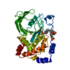 1aaxS C: citing same article ( S: Starting model for refinement |
|---|---|
| Similar structure data |
-Links
 PDBj
PDBj


- Assembly
| Deposited unit | 
| ||||||||
|---|---|---|---|---|---|---|---|---|---|
| 1 |
| ||||||||
| Unit cell |
|
-Components
| #1: Protein | Mass: 37349.574 Da / Num. of mol.: 1 / Fragment: RESIDUES 1-321 / Mutation: C215S Source method: isolated from a genetically manipulated source Source: (gene. exp.) Homo sapiens (human) / Plasmid: PUC118-PTP1B-C215S / Species (production host): Escherichia coli / Production host:  | ||
|---|---|---|---|
| #2: Protein/peptide | Mass: 1022.044 Da / Num. of mol.: 1 / Source method: obtained synthetically / Details: CHEMICALLY SYNTHESIZED | ||
| #3: Chemical | ChemComp-MG /   Mass: 24.305 Da / Num. of mol.: 1 / Source method: obtained synthetically / Formula: Mg Mass: 24.305 Da / Num. of mol.: 1 / Source method: obtained synthetically / Formula: Mg | ||
| #4: Chemical |   Mass: 60.052 Da / Num. of mol.: 2 / Source method: obtained synthetically / Formula: C2H4O2 Mass: 60.052 Da / Num. of mol.: 2 / Source method: obtained synthetically / Formula: C2H4O2#5: Water | ChemComp-HOH / |  Mass: 18.015 Da / Num. of mol.: 300 / Source method: isolated from a natural source / Formula: H2O Mass: 18.015 Da / Num. of mol.: 300 / Source method: isolated from a natural source / Formula: H2O |
-Experimental details
-Experiment
| Experiment | Method: X-RAY DIFFRACTION / Number of used crystals: 1 |
|---|
- Sample preparation
| Crystal | Density Matthews: 2.77 Å3/Da / Density % sol: 55.64 % | ||||||||||||||||||||||||||||||||||||||||||||||||||||||||||||
|---|---|---|---|---|---|---|---|---|---|---|---|---|---|---|---|---|---|---|---|---|---|---|---|---|---|---|---|---|---|---|---|---|---|---|---|---|---|---|---|---|---|---|---|---|---|---|---|---|---|---|---|---|---|---|---|---|---|---|---|---|---|
| Crystal grow | pH: 7.5 / Details: pH 7.5 | ||||||||||||||||||||||||||||||||||||||||||||||||||||||||||||
| Crystal grow | *PLUS Temperature: 4 ℃ / pH: 6.5 / Method: vapor diffusion, hanging drop | ||||||||||||||||||||||||||||||||||||||||||||||||||||||||||||
| Components of the solutions | *PLUS
|
-Data collection
| Diffraction | Mean temperature: 140 K |
|---|---|
| Diffraction source | Source: SYNCHROTRON / Site: NSLS  / Beamline: X9B / Wavelength: 1.2 / Beamline: X9B / Wavelength: 1.2 |
| Detector | Type: FUJI / Detector: IMAGE PLATE / Date: May 1, 1996 |
| Radiation | Protocol: SINGLE WAVELENGTH / Monochromatic (M) / Laue (L): M / Scattering type: x-ray |
| Radiation wavelength | Wavelength: 1.2 Å / Relative weight: 1 |
| Reflection | Resolution: 1.9→90 Å / Num. obs: 32272 / % possible obs: 93.3 % / Observed criterion σ(I): 0 / Biso Wilson estimate: 16.1 Å2 / Rmerge(I) obs: 0.035 / Net I/σ(I): 20.7 |
| Reflection shell | Resolution: 1.9→1.97 Å / Rmerge(I) obs: 0.131 / Mean I/σ(I) obs: 4.8 / % possible all: 63.7 |
| Reflection | *PLUS Num. measured all: 160542 |
| Reflection shell | *PLUS % possible obs: 63.7 % / Num. unique obs: 2160 |
- Processing
| Software |
| ||||||||||||||||||||||||||||||||||||||||||||||||||||||||||||||||||||||||||||||||
|---|---|---|---|---|---|---|---|---|---|---|---|---|---|---|---|---|---|---|---|---|---|---|---|---|---|---|---|---|---|---|---|---|---|---|---|---|---|---|---|---|---|---|---|---|---|---|---|---|---|---|---|---|---|---|---|---|---|---|---|---|---|---|---|---|---|---|---|---|---|---|---|---|---|---|---|---|---|---|---|---|---|
| Refinement | Method to determine structure: MOLECULAR REPLACEMENT Starting model: PDB ENTRY 1AAX Resolution: 1.9→19.78 Å / Rfactor Rfree error: 0.005 / Data cutoff high absF: 414834.32 / Data cutoff low absF: 0 / Isotropic thermal model: RESTRAINED / Cross valid method: THROUGHOUT / σ(I): 0 / Stereochemistry target values: Engh & Huber / Details: ONLY 8 OUT OF TWELVE PEPTIDE RESIDUES ARE VISIBLE
| ||||||||||||||||||||||||||||||||||||||||||||||||||||||||||||||||||||||||||||||||
| Solvent computation | Solvent model: FLAT MODEL / Bsol: 60.02 Å2 / ksol: 0.403 e/Å3 | ||||||||||||||||||||||||||||||||||||||||||||||||||||||||||||||||||||||||||||||||
| Displacement parameters | Biso mean: 34.4 Å2
| ||||||||||||||||||||||||||||||||||||||||||||||||||||||||||||||||||||||||||||||||
| Refine analyze |
| ||||||||||||||||||||||||||||||||||||||||||||||||||||||||||||||||||||||||||||||||
| Refinement step | Cycle: LAST / Resolution: 1.9→19.78 Å
| ||||||||||||||||||||||||||||||||||||||||||||||||||||||||||||||||||||||||||||||||
| Refine LS restraints |
| ||||||||||||||||||||||||||||||||||||||||||||||||||||||||||||||||||||||||||||||||
| LS refinement shell | Resolution: 1.9→1.97 Å / Rfactor Rfree error: 0.034 / Total num. of bins used: 10
| ||||||||||||||||||||||||||||||||||||||||||||||||||||||||||||||||||||||||||||||||
| Xplor file |
| ||||||||||||||||||||||||||||||||||||||||||||||||||||||||||||||||||||||||||||||||
| Software | *PLUS Name: CNS / Version: 0.9 / Classification: refinement | ||||||||||||||||||||||||||||||||||||||||||||||||||||||||||||||||||||||||||||||||
| Refinement | *PLUS % reflection Rfree: 5.1 % | ||||||||||||||||||||||||||||||||||||||||||||||||||||||||||||||||||||||||||||||||
| Solvent computation | *PLUS | ||||||||||||||||||||||||||||||||||||||||||||||||||||||||||||||||||||||||||||||||
| Displacement parameters | *PLUS Biso mean: 34.4 Å2 | ||||||||||||||||||||||||||||||||||||||||||||||||||||||||||||||||||||||||||||||||
| Refine LS restraints | *PLUS
| ||||||||||||||||||||||||||||||||||||||||||||||||||||||||||||||||||||||||||||||||
| LS refinement shell | *PLUS Rfactor Rfree: 0.364 / % reflection Rfree: 5.2 % / Rfactor Rwork: 0.282 / Rfactor obs: 0.282 |
