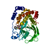
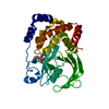
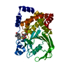
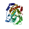
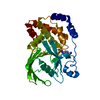
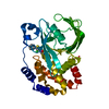
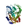
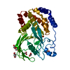
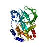
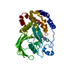
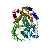
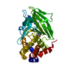
Entry Database : PDB / ID : 7l0hTitle Vanadate-bound PTP1B T177G Tyrosine-protein phosphatase non-receptor type 1 Keywords / Function / homology Function Domain/homology Component
/ / / / / / / / / / / / / / / / / / / / / / / / / / / / / / / / / / / / / / / / / / / / / / / / / / / / / / / / / / / / / / / / / / / / / / / / / / / / / / / / / / / / / / / / / / / / / / / / / / / / / / / / / / Biological species Homo sapiens (human)Method / / / Resolution : 2.1 Å Authors Shen, R.D. / Hengge, A.C. / Johnson, S.J. Funding support Organization Grant number Country National Institutes of Health/Office of the Director R01GM112781
Journal : Jacs Au / Year : 2021Title : Single Residue on the WPD-Loop Affects the pH Dependency of Catalysis in Protein Tyrosine Phosphatases.Authors : Shen, R. / Crean, R.M. / Johnson, S.J. / Kamerlin, S.C.L. / Hengge, A.C. History Deposition Dec 11, 2020 Deposition site / Processing site Revision 1.0 May 12, 2021 Provider / Type Revision 1.1 Aug 25, 2021 Group / Category / database_2Item _citation.country / _citation.journal_abbrev ... _citation.country / _citation.journal_abbrev / _citation.journal_id_ISSN / _citation.journal_volume / _citation.page_first / _citation.page_last / _citation.pdbx_database_id_PubMed / _citation.title / _database_2.pdbx_DOI / _database_2.pdbx_database_accession Revision 1.2 Oct 18, 2023 Group / Refinement descriptionCategory / chem_comp_bond / pdbx_initial_refinement_model
Show all Show less
 Movie
Movie Controller


 Open data
 Basic information
 Components
 Keywords
 Function and homology information
 Homo sapiens (human)
 X-RAY DIFFRACTION /
  Authors
 United States, 1items
  Citation
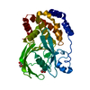
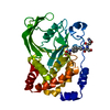
 Structure visualization
 Downloads & links
 Other downloads


 PDBj
PDBj


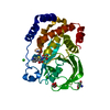
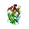
 Assembly
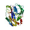


 Mass: 114.939 Da / Num. of mol.: 1 / Source method: obtained synthetically / Formula: VO4 / Feature type: SUBJECT OF INVESTIGATION
 Mass: 18.015 Da / Num. of mol.: 136 / Source method: isolated from a natural source / Formula: H2O
 Sample preparation
 Processing