 Movie
Movie Controller


+ Open data

- Basic information
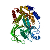
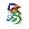
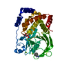
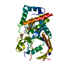
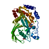
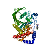
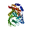
| Entry | Database: PDB / ID: 6xee | ||||||
|---|---|---|---|---|---|---|---|
| Title | Crystal Structure of the PTP1B YopH WPD loop Chimera 4 apo form | ||||||
 Components Components | Tyrosine-protein phosphatase non-receptor type 1 | ||||||
 Keywords Keywords | HYDROLASE / PTP1B / tyrosine phosphatase / WPD loop | ||||||
| Function / homology |  Function and homology information Function and homology informationPTK6 Down-Regulation / regulation of hepatocyte growth factor receptor signaling pathway / positive regulation of receptor catabolic process / insulin receptor recycling / negative regulation of vascular endothelial growth factor receptor signaling pathway / regulation of intracellular protein transport / negative regulation of MAP kinase activity / mitochondrial crista / IRE1-mediated unfolded protein response / platelet-derived growth factor receptor-beta signaling pathway ...PTK6 Down-Regulation / regulation of hepatocyte growth factor receptor signaling pathway / positive regulation of receptor catabolic process / insulin receptor recycling / negative regulation of vascular endothelial growth factor receptor signaling pathway / regulation of intracellular protein transport / negative regulation of MAP kinase activity / mitochondrial crista / IRE1-mediated unfolded protein response / platelet-derived growth factor receptor-beta signaling pathway / sorting endosome / positive regulation of IRE1-mediated unfolded protein response / cytoplasmic side of endoplasmic reticulum membrane / negative regulation of PERK-mediated unfolded protein response / regulation of type I interferon-mediated signaling pathway / negative regulation of vascular associated smooth muscle cell migration / vascular endothelial cell response to oscillatory fluid shear stress / peptidyl-tyrosine dephosphorylation / positive regulation of systemic arterial blood pressure / non-membrane spanning protein tyrosine phosphatase activity / regulation of endocytosis / Regulation of IFNA/IFNB signaling / cellular response to angiotensin / regulation of proteolysis / growth hormone receptor signaling pathway via JAK-STAT / negative regulation of cell-substrate adhesion / cellular response to unfolded protein / regulation of postsynapse assembly / positive regulation of endothelial cell apoptotic process / regulation of signal transduction / negative regulation of signal transduction / Regulation of IFNG signaling / Growth hormone receptor signaling / negative regulation of endoplasmic reticulum stress-induced intrinsic apoptotic signaling pathway / positive regulation of heart rate / ephrin receptor binding / Insulin receptor recycling / MECP2 regulates neuronal receptors and channels / cellular response to platelet-derived growth factor stimulus / Integrin signaling / endoplasmic reticulum unfolded protein response / phosphoprotein phosphatase activity / protein-tyrosine-phosphatase / cellular response to fibroblast growth factor stimulus / cellular response to nitric oxide / negative regulation of insulin receptor signaling pathway / protein tyrosine phosphatase activity / positive regulation of cardiac muscle cell apoptotic process / protein phosphatase 2A binding / Turbulent (oscillatory, disturbed) flow shear stress activates signaling by PIEZO1 and integrins in endothelial cells / endosome lumen / negative regulation of phosphatidylinositol 3-kinase/protein kinase B signal transduction / insulin receptor binding / cellular response to nerve growth factor stimulus / response to nutrient levels / negative regulation of ERK1 and ERK2 cascade / Negative regulation of MET activity / receptor tyrosine kinase binding / positive regulation of JNK cascade / insulin receptor signaling pathway / negative regulation of neuron projection development / actin cytoskeleton organization / cellular response to hypoxia / early endosome / postsynapse / cadherin binding / mitochondrial matrix / negative regulation of cell population proliferation / protein kinase binding / glutamatergic synapse / enzyme binding / endoplasmic reticulum / protein-containing complex / RNA binding / zinc ion binding / cytoplasm / cytosol Similarity search - Function | ||||||
| Biological species |  Homo sapiens (human) Homo sapiens (human) | ||||||
| Method |  X-RAY DIFFRACTION / SYNCHROTRON / MOLECULAR REPLACEMENT / molecular replacement / Resolution: 2.501 Å X-RAY DIFFRACTION / SYNCHROTRON / MOLECULAR REPLACEMENT / molecular replacement / Resolution: 2.501 Å | ||||||
 Authors Authors | Olsen, K.J. / Shen, R. / Johnson, S.J. / Hengge, A.C. | ||||||
| Funding support |  United States, 1items United States, 1items
| ||||||
 Citation Citation | Journal: Chem Sci / Year: 2022 Title: Insights into the importance of WPD-loop sequence for activity and structure in protein tyrosine phosphatases. Authors: Shen, R. / Crean, R.M. / Olsen, K.J. / Corbella, M. / Calixto, A.R. / Richan, T. / Brandao, T.A.S. / Berry, R.D. / Tolman, A. / Loria, J.P. / Johnson, S.J. / Kamerlin, S.C.L. / Hengge, A.C. | ||||||
| History |
|
- Structure visualization
| Structure viewer | Molecule: MolmilJmol/JSmol |
|---|
- Downloads & links
-Download
| PDBx/mmCIF format | 6xee.cif.gz | 78.2 KB | Display | PDBx/mmCIF format |
|---|---|---|---|---|
| PDB format | pdb6xee.ent.gz | 55 KB | Display | PDB format |
| PDBx/mmJSON format | 6xee.json.gz | Tree view | PDBx/mmJSON format | |
| Others |  Other downloads Other downloads |
-Validation report
| Arichive directory | https://data.pdbj.org/pub/pdb/validation_reports/xe/6xeeftp://data.pdbj.org/pub/pdb/validation_reports/xe/6xee | HTTPS FTP |
|---|
-Related structure data
| Related structure data | 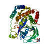 6xe8C  6xeaC  6xedC  6xefC  6xegC  7s4fC 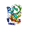 3qkpS S: Starting model for refinement C: citing same article ( |
|---|---|
| Similar structure data |


-Links
 PDBj
PDBj


- Assembly
| Deposited unit | 
| ||||||||
|---|---|---|---|---|---|---|---|---|---|
| 1 |
| ||||||||
| Unit cell |
|
-Components
| #1: Protein | Mass: 37375.590 Da / Num. of mol.: 1 Source method: isolated from a genetically manipulated source Source: (gene. exp.) Homo sapiens (human) / Gene: PTPN1, PTP1B / Production host:  | ||||||||
|---|---|---|---|---|---|---|---|---|---|
| #2: Chemical |   Mass: 120.152 Da / Num. of mol.: 2 / Source method: obtained synthetically / Formula: C7H8N2 Mass: 120.152 Da / Num. of mol.: 2 / Source method: obtained synthetically / Formula: C7H8N2#3: Chemical | ChemComp-PEG / |   Mass: 106.120 Da / Num. of mol.: 1 / Source method: obtained synthetically / Formula: C4H10O3 Mass: 106.120 Da / Num. of mol.: 1 / Source method: obtained synthetically / Formula: C4H10O3#4: Chemical | ChemComp-TRS / |   Mass: 122.143 Da / Num. of mol.: 1 / Source method: obtained synthetically / Formula: C4H12NO3 / Comment: pH buffer*YM Mass: 122.143 Da / Num. of mol.: 1 / Source method: obtained synthetically / Formula: C4H12NO3 / Comment: pH buffer*YM#5: Water | ChemComp-HOH / |  Mass: 18.015 Da / Num. of mol.: 87 / Source method: isolated from a natural source / Formula: H2O Mass: 18.015 Da / Num. of mol.: 87 / Source method: isolated from a natural source / Formula: H2OHas ligand of interest | N | |
-Experimental details
-Experiment
| Experiment | Method: X-RAY DIFFRACTION / Number of used crystals: 1 |
|---|
- Sample preparation
| Crystal | Density Matthews: 3.25 Å3/Da / Density % sol: 60.79 % |
|---|---|
| Crystal grow | Temperature: 295 K / Method: vapor diffusion, hanging drop / pH: 7.8 Details: tris hydrochloride pH 7.8, magnesium acetate tetrahydrate, PEG 8000 and benzamidine PH range: 7.8-8.5 |
-Data collection
| Diffraction | Mean temperature: 100 K / Serial crystal experiment: N | |||||||||||||||||||||||||||||||||||||||||||||||||||||||||||||||||||||||||||||||||||||||||||||||||||
|---|---|---|---|---|---|---|---|---|---|---|---|---|---|---|---|---|---|---|---|---|---|---|---|---|---|---|---|---|---|---|---|---|---|---|---|---|---|---|---|---|---|---|---|---|---|---|---|---|---|---|---|---|---|---|---|---|---|---|---|---|---|---|---|---|---|---|---|---|---|---|---|---|---|---|---|---|---|---|---|---|---|---|---|---|---|---|---|---|---|---|---|---|---|---|---|---|---|---|---|---|
| Diffraction source | Source: SYNCHROTRON / Site: SSRL / Beamline: BL9-1 / Wavelength: 0.97946 Å | |||||||||||||||||||||||||||||||||||||||||||||||||||||||||||||||||||||||||||||||||||||||||||||||||||
| Detector | Type: ADSC QUANTUM 315r / Detector: CCD / Date: Jan 31, 2019 | |||||||||||||||||||||||||||||||||||||||||||||||||||||||||||||||||||||||||||||||||||||||||||||||||||
| Radiation | Protocol: SINGLE WAVELENGTH / Monochromatic (M) / Laue (L): M / Scattering type: x-ray | |||||||||||||||||||||||||||||||||||||||||||||||||||||||||||||||||||||||||||||||||||||||||||||||||||
| Radiation wavelength | Wavelength: 0.97946 Å / Relative weight: 1 | |||||||||||||||||||||||||||||||||||||||||||||||||||||||||||||||||||||||||||||||||||||||||||||||||||
| Reflection | Resolution: 2.5→50 Å / Num. obs: 16320 / % possible obs: 97.7 % / Redundancy: 16 % / Rmerge(I) obs: 0.137 / Rpim(I) all: 0.033 / Rrim(I) all: 0.142 / Χ2: 1.027 / Net I/σ(I): 7.2 / Num. measured all: 261786 | |||||||||||||||||||||||||||||||||||||||||||||||||||||||||||||||||||||||||||||||||||||||||||||||||||
| Reflection shell | Diffraction-ID: 1
|
-Phasing
| Phasing | Method: molecular replacement | |||||||||
|---|---|---|---|---|---|---|---|---|---|---|
| Phasing MR |
|
- Processing
| Software |
| ||||||||||||||||||||||||||||||||||||||||||
|---|---|---|---|---|---|---|---|---|---|---|---|---|---|---|---|---|---|---|---|---|---|---|---|---|---|---|---|---|---|---|---|---|---|---|---|---|---|---|---|---|---|---|---|
| Refinement | Method to determine structure: MOLECULAR REPLACEMENT Starting model: 3QKP Resolution: 2.501→40.676 Å / SU ML: 0.28 / Cross valid method: THROUGHOUT / σ(F): 1.35 / Phase error: 21.13 / Stereochemistry target values: ML
| ||||||||||||||||||||||||||||||||||||||||||
| Solvent computation | Shrinkage radii: 0.9 Å / VDW probe radii: 1.11 Å / Solvent model: FLAT BULK SOLVENT MODEL | ||||||||||||||||||||||||||||||||||||||||||
| Displacement parameters | Biso max: 142.56 Å2 / Biso mean: 57.299 Å2 / Biso min: 27.97 Å2 | ||||||||||||||||||||||||||||||||||||||||||
| Refinement step | Cycle: final / Resolution: 2.501→40.676 Å
| ||||||||||||||||||||||||||||||||||||||||||
| LS refinement shell | Refine-ID: X-RAY DIFFRACTION / Rfactor Rfree error: 0
|
