 Movie
Movie Controller


+ Open data

- Basic information
| Entry | Database: PDB / ID: 6bab | ||||||
|---|---|---|---|---|---|---|---|
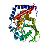
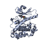
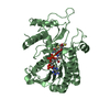
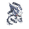
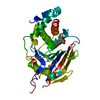
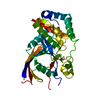
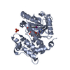
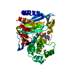
| Title | The structure of human CamKII with bound inhibitor | ||||||
 Components Components | Calcium/calmodulin-dependent protein kinase type II subunit delta | ||||||
 Keywords Keywords | TRANSFERASE/TRANSFERASE Inhibitor / Kinase / CamKII / inhibitor / TRANSFERASE / TRANSFERASE-TRANSFERASE Inhibitor complex | ||||||
| Function / homology |  Function and homology information Function and homology informationHSF1-dependent transactivation / regulation of relaxation of cardiac muscle / Interferon gamma signaling / regulation of cellular localization / Ion transport by P-type ATPases / RAF activation / cell growth involved in cardiac muscle cell development / cochlear hair cell ribbon synapse / regulation of cell communication by electrical coupling / Unblocking of NMDA receptors, glutamate binding and activation ...HSF1-dependent transactivation / regulation of relaxation of cardiac muscle / Interferon gamma signaling / regulation of cellular localization / Ion transport by P-type ATPases / RAF activation / cell growth involved in cardiac muscle cell development / cochlear hair cell ribbon synapse / regulation of cell communication by electrical coupling / Unblocking of NMDA receptors, glutamate binding and activation / Ca2+/calmodulin-dependent protein kinase / Trafficking of AMPA receptors / RAF/MAP kinase cascade / Ion homeostasis / axon initial segment / endoplasmic reticulum calcium ion homeostasis / calcium/calmodulin-dependent protein kinase activity / regulation of calcium ion transmembrane transport via high voltage-gated calcium channel / relaxation of cardiac muscle / positive regulation of smooth muscle cell migration / positive regulation of DNA biosynthetic process / cardiac muscle cell contraction / positive regulation of cardiac muscle hypertrophy / peptidyl-threonine phosphorylation / regulation of cardiac muscle cell action potential / intracellular potassium ion homeostasis / nitric-oxide synthase binding / intercalated disc / positive regulation of G2/M transition of mitotic cell cycle / postsynaptic cytosol / positive regulation of Rac protein signal transduction / positive regulation of vascular associated smooth muscle cell proliferation / regulation of protein localization to plasma membrane / regulation of sodium ion transport / regulation of release of sequestered calcium ion into cytosol by sarcoplasmic reticulum / cardiac muscle contraction / positive regulation of cardiac muscle cell apoptotic process / sarcoplasmic reticulum membrane / T-tubule / neuromuscular junction / G1/S transition of mitotic cell cycle / calcium ion transport / protein phosphorylation / transmembrane transporter binding / response to hypoxia / positive regulation of ERK1 and ERK2 cascade / protein kinase activity / calmodulin binding / neuron projection / protein serine kinase activity / protein serine/threonine kinase activity / neuronal cell body / perinuclear region of cytoplasm / protein-containing complex / ATP binding / nucleus / cytoplasm Similarity search - Function | ||||||
| Biological species |  | ||||||
| Method |  X-RAY DIFFRACTION / SYNCHROTRON / MOLECULAR REPLACEMENT / Resolution: 1.91 Å X-RAY DIFFRACTION / SYNCHROTRON / MOLECULAR REPLACEMENT / Resolution: 1.91 Å | ||||||
 Authors Authors | Somoza, J.R. / Villasenor, A.G. | ||||||
 Citation Citation | Journal: To Be Published Title: TBD Authors: Koltun, D. / Somoza, J.R. | ||||||
| History |
|
- Structure visualization
| Structure viewer | Molecule: MolmilJmol/JSmol |
|---|
- Downloads & links
-Download
| PDBx/mmCIF format | 6bab.cif.gz | 261.6 KB | Display | PDBx/mmCIF format |
|---|---|---|---|---|
| PDB format | pdb6bab.ent.gz | 210.6 KB | Display | PDB format |
| PDBx/mmJSON format | 6bab.json.gz | Tree view | PDBx/mmJSON format | |
| Others |  Other downloads Other downloads |
-Validation report
| Arichive directory | https://data.pdbj.org/pub/pdb/validation_reports/ba/6babftp://data.pdbj.org/pub/pdb/validation_reports/ba/6bab | HTTPS FTP |
|---|
-Related structure data
| Similar structure data |
|---|


-Links
 PDBj
PDBj


- Assembly
| Deposited unit | 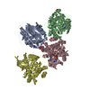
| ||||||||||
|---|---|---|---|---|---|---|---|---|---|---|---|
| 1 | 
| ||||||||||
| 2 | 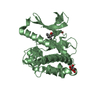
| ||||||||||
| 3 | 
| ||||||||||
| 4 | 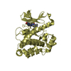
| ||||||||||
| Unit cell |
|
-Components
| #1: Protein | Mass: 34459.574 Da / Num. of mol.: 4 Source method: isolated from a genetically manipulated source Source: (gene. exp.)   Spodoptera frugiperda (fall armyworm) Spodoptera frugiperda (fall armyworm)References: UniProt: Q6PHZ2, Ca2+/calmodulin-dependent protein kinase #2: Chemical | ChemComp-D0S /   Mass: 505.635 Da / Num. of mol.: 4 / Source method: obtained synthetically / Formula: C26H31N7O2S Mass: 505.635 Da / Num. of mol.: 4 / Source method: obtained synthetically / Formula: C26H31N7O2S#3: Chemical |   Mass: 150.087 Da / Num. of mol.: 3 / Source method: obtained synthetically / Formula: C4H6O6 Mass: 150.087 Da / Num. of mol.: 3 / Source method: obtained synthetically / Formula: C4H6O6#4: Water | ChemComp-HOH / |  Mass: 18.015 Da / Num. of mol.: 936 / Source method: isolated from a natural source / Formula: H2O Mass: 18.015 Da / Num. of mol.: 936 / Source method: isolated from a natural source / Formula: H2OHas protein modification | Y | |
|---|
-Experimental details
-Experiment
| Experiment | Method: X-RAY DIFFRACTION / Number of used crystals: 1 |
|---|
- Sample preparation
| Crystal | Density Matthews: 2.3 Å3/Da / Density % sol: 46.58 % |
|---|---|
| Crystal grow | Temperature: 293 K / Method: vapor diffusion Details: Sitting drop vapor diffusion droplets were assembled with 250 nL of 12 mg/mL CamKII, 0.6 mM inhibitor and 250 nL of reservoir solution 24% peg 3350, 0.2 M ammonium tartrate, 0.1 M arginine. |
-Data collection
| Diffraction | Mean temperature: 100 K |
|---|---|
| Diffraction source | Source: SYNCHROTRON / Site: APS  / Beamline: 21-ID-D / Wavelength: 1.08 Å / Beamline: 21-ID-D / Wavelength: 1.08 Å |
| Detector | Type: DECTRIS EIGER X 9M / Detector: PIXEL / Date: Feb 21, 2017 |
| Radiation | Protocol: SINGLE WAVELENGTH / Monochromatic (M) / Laue (L): M / Scattering type: x-ray |
| Radiation wavelength | Wavelength: 1.08 Å / Relative weight: 1 |
| Reflection | Resolution: 1.91→54.2 Å / Num. obs: 80037 / % possible obs: 87.4 % / Redundancy: 2 % / Biso Wilson estimate: 22.25 Å2 / Net I/σ(I): 7.6 |
- Processing
| Software |
| |||||||||||||||||||||||||||||||||||||||||||||||||||||||||||||||||||||||||||||
|---|---|---|---|---|---|---|---|---|---|---|---|---|---|---|---|---|---|---|---|---|---|---|---|---|---|---|---|---|---|---|---|---|---|---|---|---|---|---|---|---|---|---|---|---|---|---|---|---|---|---|---|---|---|---|---|---|---|---|---|---|---|---|---|---|---|---|---|---|---|---|---|---|---|---|---|---|---|---|
| Refinement | Method to determine structure: MOLECULAR REPLACEMENT / Resolution: 1.91→54.18 Å / SU ML: 0.248 / Cross valid method: FREE R-VALUE / σ(F): 1.986 / Phase error: 27.233 Stereochemistry target values: GEOSTD + MONOMER LIBRARY + CDL V1.2
| |||||||||||||||||||||||||||||||||||||||||||||||||||||||||||||||||||||||||||||
| Solvent computation | Shrinkage radii: 0.9 Å / VDW probe radii: 1.11 Å / Solvent model: FLAT BULK SOLVENT MODEL | |||||||||||||||||||||||||||||||||||||||||||||||||||||||||||||||||||||||||||||
| Displacement parameters | Biso mean: 25.33 Å2 | |||||||||||||||||||||||||||||||||||||||||||||||||||||||||||||||||||||||||||||
| Refinement step | Cycle: LAST / Resolution: 1.91→54.18 Å
| |||||||||||||||||||||||||||||||||||||||||||||||||||||||||||||||||||||||||||||
| Refine LS restraints |
| |||||||||||||||||||||||||||||||||||||||||||||||||||||||||||||||||||||||||||||
| LS refinement shell |
|
