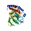
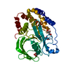
Entry Database : PDB / ID : 6krwTitle Crystal Structure of AtPTP1 at 1.4 angstrom Protein-tyrosine-phosphatase PTP1 Keywords / / Function / homology Function Domain/homology Component
/ / / / / / / / / / / / / / / / / / / / / / / / / / / / / / / / / Biological species Arabidopsis thaliana (thale cress)Method / / / Resolution : 1.4 Å Authors Zhao, Y.Y. / Luo, Z.P. / Wang, J. / Wu, J.W. Funding support Organization Grant number Country National Science Foundation (China)
Journal : To Be Published Title : Crystal structure of AtPTP1 at 1.4 AngstromsAuthors : Zhao, Y.Y. / Wang, H.J. / Hu, J. / Tang, J. / Zhang, W.H. / He, Q.Q. / Deng, H.T. / Luo, Z.P. / Wang, J. / Wang, Z.X. / Wang, X.L. / Wu, J.W. History Deposition Aug 22, 2019 Deposition site / Processing site Revision 1.0 Aug 26, 2020 Provider / Type Revision 1.1 Nov 22, 2023 Group / Database references / Refinement descriptionCategory chem_comp_atom / chem_comp_bond ... chem_comp_atom / chem_comp_bond / database_2 / pdbx_initial_refinement_model Item / _database_2.pdbx_database_accession
Show all Show less
 Movie
Movie Controller


 Open data
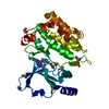
 Basic information
 Components
 Keywords
 Function and homology information
 X-RAY DIFFRACTION /
  Authors
 China, 1items
  Citation
 Structure visualization
 Downloads & links
 Other downloads
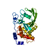


 PDBj
PDBj


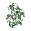
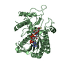
 Assembly
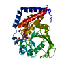


 Mass: 126.904 Da / Num. of mol.: 3 / Source method: obtained synthetically / Formula: I
 Mass: 189.100 Da / Num. of mol.: 1 / Source method: obtained synthetically / Formula: C6H5O7
 Mass: 106.120 Da / Num. of mol.: 1 / Source method: obtained synthetically / Formula: C4H10O3
 Mass: 18.015 Da / Num. of mol.: 196 / Source method: isolated from a natural source / Formula: H2O
 Sample preparation
 Processing