Resolution: 1.7→37.201 Å / Cor.coef. Fo:Fc: 0.962 / Cor.coef. Fo:Fc free: 0.954 / SU B: 1.539 / SU ML: 0.051 / Cross valid method: THROUGHOUT / ESU R: 0.093 / ESU R Free: 0.088 Details: Hydrogens have been added in their riding positions
Rfactor
Num. reflection
% reflection
Selection details
Rfree
0.1743
1713
5.026 %
Random selection
Rwork
0.1511
-
-
-
all
0.152
-
-
-
obs
-
34081
96.465 %
-
Solvent computation
Ion probe radii: 0.8 Å / Shrinkage radii: 0.8 Å / VDW probe radii: 1.2 Å
Displacement parameters
Biso mean: 16.394 Å2
Baniso -1
Baniso -2
Baniso -3
1-
0.138 Å2
0 Å2
0.088 Å2
2-
-
-0.251 Å2
0 Å2
3-
-
-
0.035 Å2
Refinement step
Cycle: LAST / Resolution: 1.7→37.201 Å
Protein
Nucleic acid
Ligand
Solvent
Total
Num. atoms
2391
0
15
216
2622
Refine LS restraints
Refine-ID
Type
Dev ideal
Dev ideal target
Number
X-RAY DIFFRACTION
r_bond_refined_d
0.006
0.013
2517
X-RAY DIFFRACTION
r_bond_other_d
0.001
0.017
2298
X-RAY DIFFRACTION
r_angle_refined_deg
1.279
1.641
3428
X-RAY DIFFRACTION
r_angle_other_deg
0.339
1.571
5358
X-RAY DIFFRACTION
r_dihedral_angle_1_deg
6.105
5
315
X-RAY DIFFRACTION
r_dihedral_angle_2_deg
32.11
22.977
131
X-RAY DIFFRACTION
r_dihedral_angle_3_deg
11.811
15
441
X-RAY DIFFRACTION
r_dihedral_angle_4_deg
13.933
15
15
X-RAY DIFFRACTION
r_chiral_restr
0.057
0.2
337
X-RAY DIFFRACTION
r_gen_planes_refined
0.056
0.02
2834
X-RAY DIFFRACTION
r_gen_planes_other
0.052
0.02
503
X-RAY DIFFRACTION
r_nbd_refined
0.211
0.2
500
X-RAY DIFFRACTION
r_symmetry_nbd_other
0.163
0.2
2230
X-RAY DIFFRACTION
r_nbtor_refined
0.167
0.2
1256
X-RAY DIFFRACTION
r_symmetry_nbtor_other
0.091
0.2
1105
X-RAY DIFFRACTION
r_xyhbond_nbd_refined
0.149
0.2
142
X-RAY DIFFRACTION
r_symmetry_xyhbond_nbd_other
0.107
0.2
1
X-RAY DIFFRACTION
r_symmetry_nbd_refined
0.136
0.2
15
X-RAY DIFFRACTION
r_nbd_other
0.166
0.2
55
X-RAY DIFFRACTION
r_symmetry_xyhbond_nbd_refined
0.154
0.2
14
X-RAY DIFFRACTION
r_xyhbond_nbd_other
0.087
0.2
1
X-RAY DIFFRACTION
r_mcbond_it
2.334
3.024
1242
X-RAY DIFFRACTION
r_mcbond_other
2.328
3.021
1241
X-RAY DIFFRACTION
r_mcangle_it
3.554
5.657
1563
X-RAY DIFFRACTION
r_mcangle_other
3.556
5.66
1564
X-RAY DIFFRACTION
r_scbond_it
3.292
3.715
1275
X-RAY DIFFRACTION
r_scbond_other
3.29
3.714
1274
X-RAY DIFFRACTION
r_scangle_it
5.041
6.615
1865
X-RAY DIFFRACTION
r_scangle_other
5.04
6.615
1866
X-RAY DIFFRACTION
r_lrange_it
6.357
31.959
10915
X-RAY DIFFRACTION
r_lrange_other
6.271
31.69
10783
LS refinement shell
Resolution (Å)
Rfactor Rfree
Num. reflection Rfree
Rfactor Rwork
Num. reflection Rwork
Refine-ID
% reflection obs (%)
1.7-1.743
0.198
82
0.182
1815
X-RAY DIFFRACTION
74.9802
1.743-1.79
0.198
111
0.163
2159
X-RAY DIFFRACTION
90.1151
1.79-1.842
0.173
128
0.15
2332
X-RAY DIFFRACTION
99.3939
1.842-1.899
0.164
123
0.149
2231
X-RAY DIFFRACTION
99.4088
1.899-1.961
0.175
131
0.154
2164
X-RAY DIFFRACTION
98.5401
1.961-2.029
0.171
117
0.147
2097
X-RAY DIFFRACTION
99.1047
2.029-2.106
0.179
120
0.148
1979
X-RAY DIFFRACTION
96.9963
2.106-2.192
0.148
131
0.136
1952
X-RAY DIFFRACTION
99.5222
2.192-2.289
0.192
91
0.143
1908
X-RAY DIFFRACTION
99.2552
2.289-2.4
0.186
86
0.136
1775
X-RAY DIFFRACTION
99.2004
2.4-2.529
0.165
105
0.135
1682
X-RAY DIFFRACTION
98.0252
2.529-2.682
0.179
91
0.138
1634
X-RAY DIFFRACTION
99.0241
2.682-2.866
0.186
70
0.146
1537
X-RAY DIFFRACTION
99.2588
2.866-3.094
0.165
63
0.148
1457
X-RAY DIFFRACTION
99.0228
3.094-3.387
0.177
63
0.15
1311
X-RAY DIFFRACTION
98.213
3.387-3.783
0.186
52
0.158
1217
X-RAY DIFFRACTION
98.986
3.783-4.36
0.159
57
0.152
1068
X-RAY DIFFRACTION
98.8577
4.36-5.322
0.167
43
0.149
896
X-RAY DIFFRACTION
97.8125
5.322-7.451
0.169
29
0.212
729
X-RAY DIFFRACTION
98.6979
7.451-37.201
0.193
20
0.183
425
X-RAY DIFFRACTION
96.9499
+
About Yorodumi
-
News
-
Feb 9, 2022. New format data for meta-information of EMDB entries
New format data for meta-information of EMDB entries
Version 3 of the EMDB header file is now the official format.
The previous official version 1.9 will be removed from the archive.
In the structure databanks used in Yorodumi, some data are registered as the other names, "COVID-19 virus" and "2019-nCoV". Here are the details of the virus and the list of structure data.
Jan 31, 2019. EMDB accession codes are about to change! (news from PDBe EMDB page)
EMDB accession codes are about to change! (news from PDBe EMDB page)
The allocation of 4 digits for EMDB accession codes will soon come to an end. Whilst these codes will remain in use, new EMDB accession codes will include an additional digit and will expand incrementally as the available range of codes is exhausted. The current 4-digit format prefixed with “EMD-” (i.e. EMD-XXXX) will advance to a 5-digit format (i.e. EMD-XXXXX), and so on. It is currently estimated that the 4-digit codes will be depleted around Spring 2019, at which point the 5-digit format will come into force.
The EM Navigator/Yorodumi systems omit the EMD- prefix.
Related info.:Q: What is EMD? / ID/Accession-code notation in Yorodumi/EM Navigator
Yorodumi is a browser for structure data from EMDB, PDB, SASBDB, etc.
This page is also the successor to EM Navigator detail page, and also detail information page/front-end page for Omokage search.
The word "yorodu" (or yorozu) is an old Japanese word meaning "ten thousand". "mi" (miru) is to see.
Related info.:EMDB / PDB / SASBDB / Comparison of 3 databanks / Yorodumi Search / Aug 31, 2016. New EM Navigator & Yorodumi / Yorodumi Papers / Jmol/JSmol / Function and homology information / Changes in new EM Navigator and Yorodumi
 Movie
Movie Controller


 Open data
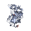
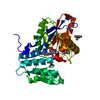
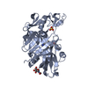
 Basic information
 Components
 Keywords
 Function and homology information
 X-RAY DIFFRACTION /
  Authors
 China, 1items
  Citation
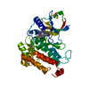
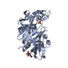
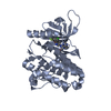
 Structure visualization
 Downloads & links
 Other downloads
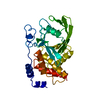
 PDBj
PDBj


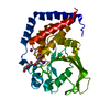
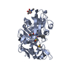
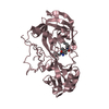
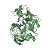
 Assembly
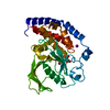


 Mass: 126.904 Da / Num. of mol.: 2 / Source method: obtained synthetically / Formula: I
 Mass: 189.100 Da / Num. of mol.: 1 / Source method: obtained synthetically / Formula: C6H5O7
 Mass: 18.015 Da / Num. of mol.: 216 / Source method: isolated from a natural source / Formula: H2O
 Sample preparation
 Processing