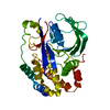
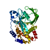
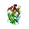
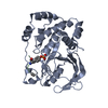
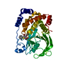
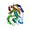
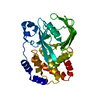
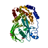
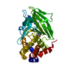
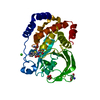
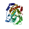
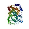
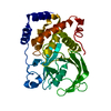
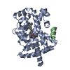
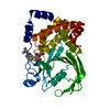
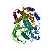
[English] 日本語
Yorodumi
- PDB-1nwe: Ptp1B R47C Modified at C47 with N-[4-(2-{2-[3-(2-Bromo-acetylamin... -

+
Open data


ID or keywords:

Loading...

-
Basic information

Entry
Database: PDB / ID: 1nwe
TitlePtp1B R47C Modified at C47 with N-[4-(2-{2-[3-(2-Bromo-acetylamino)-propionylamino]-3-hydroxy-propionylamino}-ethyl)-phenyl]-oxalamic acid
ComponentsProtein-tyrosine phosphatase, non-receptor type 1
KeywordsHYDROLASE / Acetylation / Phosphorylation / phospatase / phosphotyrosine mimetic
Function / homology
Function and homology information


regulation of hepatocyte growth factor receptor signaling pathway / PTK6 Down-Regulation / peptidyl-tyrosine dephosphorylation involved in inactivation of protein kinase activity / positive regulation of receptor catabolic process / insulin receptor recycling / negative regulation of PERK-mediated unfolded protein response / negative regulation of vascular endothelial growth factor receptor signaling pathway / regulation of intracellular protein transport / IRE1-mediated unfolded protein response / cytoplasmic side of endoplasmic reticulum membrane ...regulation of hepatocyte growth factor receptor signaling pathway / PTK6 Down-Regulation / peptidyl-tyrosine dephosphorylation involved in inactivation of protein kinase activity / positive regulation of receptor catabolic process / insulin receptor recycling / negative regulation of PERK-mediated unfolded protein response / negative regulation of vascular endothelial growth factor receptor signaling pathway / regulation of intracellular protein transport / IRE1-mediated unfolded protein response / cytoplasmic side of endoplasmic reticulum membrane / platelet-derived growth factor receptor-beta signaling pathway / sorting endosome / mitochondrial crista / positive regulation of IRE1-mediated unfolded protein response / regulation of type I interferon-mediated signaling pathway / regulation of endocytosis / non-membrane spanning protein tyrosine phosphatase activity / positive regulation of protein tyrosine kinase activity / peptidyl-tyrosine dephosphorylation / Regulation of IFNA/IFNB signaling / regulation of signal transduction / cellular response to unfolded protein / growth hormone receptor signaling pathway via JAK-STAT / negative regulation of endoplasmic reticulum stress-induced intrinsic apoptotic signaling pathway / negative regulation of signal transduction / Regulation of IFNG signaling / Growth hormone receptor signaling / MECP2 regulates neuronal receptors and channels / endoplasmic reticulum unfolded protein response / positive regulation of JUN kinase activity / Insulin receptor recycling / negative regulation of insulin receptor signaling pathway / ephrin receptor binding / Integrin signaling / protein dephosphorylation / protein-tyrosine-phosphatase / negative regulation of MAP kinase activity / protein phosphatase 2A binding / protein tyrosine phosphatase activity / endosome lumen / insulin receptor binding / Negative regulation of MET activity / negative regulation of ERK1 and ERK2 cascade / receptor tyrosine kinase binding / insulin receptor signaling pathway / actin cytoskeleton organization / early endosome / mitochondrial matrix / cadherin binding / protein kinase binding / enzyme binding / endoplasmic reticulum / protein-containing complex / RNA binding / zinc ion binding / cytoplasm / cytosol
Similarity search - Function
Protein-tyrosine phosphatase, non-receptor type-1/2 / Protein tyrosine phosphatase superfamily / Protein-Tyrosine Phosphatase; Chain A / Protein tyrosine phosphatase, catalytic domain / PTP type protein phosphatase domain profile. / Protein-tyrosine phosphatase / Tyrosine-specific protein phosphatase, PTPase domain / Protein-tyrosine phosphatase, catalytic / Protein tyrosine phosphatase, catalytic domain motif / Tyrosine specific protein phosphatases active site. ...Protein-tyrosine phosphatase, non-receptor type-1/2 / Protein tyrosine phosphatase superfamily / Protein-Tyrosine Phosphatase; Chain A / Protein tyrosine phosphatase, catalytic domain / PTP type protein phosphatase domain profile. / Protein-tyrosine phosphatase / Tyrosine-specific protein phosphatase, PTPase domain / Protein-tyrosine phosphatase, catalytic / Protein tyrosine phosphatase, catalytic domain motif / Tyrosine specific protein phosphatases active site. / Protein-tyrosine phosphatase, active site / Tyrosine-specific protein phosphatases domain / Tyrosine specific protein phosphatases domain profile. / Protein-tyrosine phosphatase-like / Alpha-Beta Complex / Alpha Beta
Similarity search - Domain/homology
Chem-FG1 / Tyrosine-protein phosphatase non-receptor type 1
Similarity search - Component
Biological speciesHomo sapiens (human)
MethodX-RAY DIFFRACTION / FOURIER SYNTHESIS / Resolution: 3.1 Å
AuthorsErlanson, D.A. / McDowell, R.S. / He, M.M. / Randal, M. / Simmons, R.L. / Kung, J. / Waight, A. / Hansen, S.K.
CitationJournal: J.Am.Chem.Soc. / Year: 2003
Title: Discovery of a New Phosphotyrosine Mimetic for PTP1B Using Breakaway Tethering
Authors: Erlanson, D.A. / McDowell, R.S. / He, M.M. / Randal, M. / Simmons, R.L. / Kung, J. / Waight, A. / Hansen, S.K.
History
DepositionFeb 6, 2003Deposition site: RCSB / Processing site: RCSB
Revision 1.0May 6, 2003Provider: repository / Type: Initial release
Revision 1.1Apr 29, 2008Group: Version format compliance
Revision 1.2Jul 13, 2011Group: Version format compliance
Revision 1.3Oct 11, 2017Group: Advisory / Refinement description / Category: pdbx_unobs_or_zero_occ_atoms / software
Revision 1.4Oct 27, 2021Group: Advisory / Database references / Derived calculations
Category: database_2 / pdbx_unobs_or_zero_occ_atoms ...database_2 / pdbx_unobs_or_zero_occ_atoms / struct_conn / struct_ref_seq_dif / struct_site
Item: _database_2.pdbx_DOI / _database_2.pdbx_database_accession ..._database_2.pdbx_DOI / _database_2.pdbx_database_accession / _struct_conn.pdbx_leaving_atom_flag / _struct_ref_seq_dif.details / _struct_site.pdbx_auth_asym_id / _struct_site.pdbx_auth_comp_id / _struct_site.pdbx_auth_seq_id
Revision 1.5Apr 3, 2024Group: Data collection / Refinement description
Category: chem_comp_atom / chem_comp_bond / pdbx_initial_refinement_model
Remark 600Heterogen The Br atom of the ligand was displaced during the covalent modification of the protein.

-
Structure visualization

Structure viewerMolecule:
MolmilJmol/JSmol

Downloads & links

-
Assembly

Deposited unit
A: Protein-tyrosine phosphatase, non-receptor type 1
hetero molecules


Theoretical massNumber of molelcules
Total (without water)35,1342
Polymers34,6461
Non-polymers4871
Water00
1


  • Idetical with deposited unit
  • defined by author
TypeNameSymmetry operationNumber
identity operation1_555x,y,z1
Unit cell
Length a, b, c (Å)88.052, 88.052, 117.594
Angle α, β, γ (deg.)90.00, 90.00, 120.00
Int Tables number152
Space group name H-MP3121

-
Components

#1: Protein Protein-tyrosine phosphatase, non-receptor type 1 / Protein-tyrosine phosphatase 1B / PTP-1B


Mass: 34646.441 Da / Num. of mol.: 1 / Fragment: catalytic domain, residues 1-298 / Mutation: C32S, R47C, C92V
Source method: isolated from a genetically manipulated source
Source: (gene. exp.) Homo sapiens (human) / Gene: PTPN1 / Plasmid: pRSET / Species (production host): Escherichia coli / Production host: Escherichia coli BL21 (bacteria) / Strain (production host): BL21 / References: UniProt: P18031, protein-tyrosine-phosphatase
#2: Chemical ChemComp-FG1 / N-[4-(2-{2-[3-(2-BROMO-ACETYLAMINO)-PROPIONYLAMINO]-3-HYDROXY-PROPIONYLAMINO}-ETHYL)-PHENYL]-OXALAMIC ACID


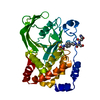
Mass: 487.302 Da / Num. of mol.: 1 / Source method: obtained synthetically / Formula: C18H23BrN4O7

-
Experimental details

-
Experiment

ExperimentMethod: X-RAY DIFFRACTION / Number of used crystals: 1

-
Sample preparation

CrystalDensity Matthews: 3.93 Å3/Da / Density % sol: 68.49 %
Crystal growTemperature: 298 K / Method: vapor diffusion, hanging drop / pH: 7.4
Details: PEG 8k, HEPES, MgOAc, pH 7.4, VAPOR DIFFUSION, HANGING DROP, temperature 298K

-
Data collection

DiffractionMean temperature: 120 K
Diffraction sourceSource: ROTATING ANODE / Type: RIGAKU / Wavelength: 1.5418 Å
DetectorType: RIGAKU RAXIS IV / Detector: IMAGE PLATE / Date: Mar 6, 2002 / Details: mirrors
RadiationMonochromator: Yale mirrors / Protocol: SINGLE WAVELENGTH / Monochromatic (M) / Laue (L): M / Scattering type: x-ray
Radiation wavelengthWavelength: 1.5418 Å / Relative weight: 1
ReflectionResolution: 3.1→15 Å / Num. all: 9867 / Num. obs: 9867 / % possible obs: 94.2 % / Observed criterion σ(F): 0 / Observed criterion σ(I): 0 / Redundancy: 2 % / Biso Wilson estimate: 47 Å2 / Rmerge(I) obs: 0.187 / Rsym value: 0.187 / Net I/σ(I): 5.8
Reflection shellResolution: 3.1→3.21 Å / Redundancy: 2 % / Rmerge(I) obs: 0.397 / Mean I/σ(I) obs: 2.6 / Num. unique all: 964 / Rsym value: 0.397 / % possible all: 94

-
Processing

Software
NameVersionClassification
REFMAC5.1.19refinement
d*TREKdata scaling
RefinementMethod to determine structure: FOURIER SYNTHESIS
Starting model: native PTP-1B

Resolution: 3.1→15 Å / Cor.coef. Fo:Fc: 0.82 / Cor.coef. Fo:Fc free: 0.808 / SU B: 35.84 / SU ML: 0.581 / Isotropic thermal model: overall / Cross valid method: THROUGHOUT / σ(F): 0 / σ(I): 0 / ESU R Free: 0.519 / Stereochemistry target values: MAXIMUM LIKELIHOOD
RfactorNum. reflection% reflectionSelection details
Rfree0.30544 447 4.9 %RANDOM
Rwork0.28295 ---
all0.284 8660 --
obs0.28401 8660 92.3 %-
Solvent computationIon probe radii: 0.8 Å / Shrinkage radii: 0.8 Å / VDW probe radii: 1.4 Å / Solvent model: BABINET MODEL WITH MASK
Displacement parametersBiso mean: 2.352 Å2
Baniso -1Baniso -2Baniso -3
1--2.6 Å2-1.3 Å20 Å2
2---2.6 Å20 Å2
3---3.9 Å2
Refinement stepCycle: LAST / Resolution: 3.1→15 Å
ProteinNucleic acidLigandSolventTotal
Num. atoms2362 0 29 0 2391
Refine LS restraints
Refine-IDTypeDev idealDev ideal targetNumber
X-RAY DIFFRACTIONr_bond_refined_d0.0080.0212442
X-RAY DIFFRACTIONr_angle_refined_deg0.9541.9553301
X-RAY DIFFRACTIONr_dihedral_angle_1_deg4.5715294
X-RAY DIFFRACTIONr_chiral_restr0.0590.2355
X-RAY DIFFRACTIONr_gen_planes_refined0.0030.021865
X-RAY DIFFRACTIONr_nbd_refined0.1980.21098
X-RAY DIFFRACTIONr_xyhbond_nbd_refined0.1160.280
X-RAY DIFFRACTIONr_symmetry_vdw_refined0.1790.217
X-RAY DIFFRACTIONr_mcbond_it0.362.51474
X-RAY DIFFRACTIONr_mcangle_it0.49652373
X-RAY DIFFRACTIONr_scbond_it0.4462.5968
X-RAY DIFFRACTIONr_scangle_it0.6245928
LS refinement shellResolution: 3.1→3.177 Å / Total num. of bins used: 20
RfactorNum. reflection% reflection
Rfree0.431 40 -
Rwork0.379 592 -
obs-632 94 %
Refinement TLS params.Method: refined / Origin x: 40.1008 Å / Origin y: 46.131 Å / Origin z: 42.4608 Å
111213212223313233
T0.0029 Å2-0.0302 Å2-0.0087 Å2-0.5577 Å2-0.0028 Å2--0.0625 Å2
L7.1211 °2-1.8696 °2-0.5645 °2-3.886 °21.1956 °2--4.0721 °2
S-0.0255 Å °0.6446 Å °0.328 Å °-0.1088 Å °-0.0216 Å °-0.1167 Å °-0.3754 Å °0.1669 Å °0.0471 Å °
Refinement TLS group
IDRefine-IDRefine TLS-IDAuth asym-IDLabel asym-IDAuth seq-IDLabel seq-ID
1X-RAY DIFFRACTION1AA2 - 2972 - 297
2X-RAY DIFFRACTION1AB10011

+
About Yorodumi

-
News

-
Feb 9, 2022. New format data for meta-information of EMDB entries

New format data for meta-information of EMDB entries

  • Version 3 of the EMDB header file is now the official format.
  • The previous official version 1.9 will be removed from the archive.

Related info.:EMDB header

External links:wwPDB to switch to version 3 of the EMDB data model

-
Aug 12, 2020. Covid-19 info

Covid-19 info

URL: https://pdbjlvh1.pdbj.org/emnavi/covid19.php

New page: Covid-19 featured information page in EM Navigator.

Related info.:Covid-19 info / Mar 5, 2020. Novel coronavirus structure data

+
Mar 5, 2020. Novel coronavirus structure data

Novel coronavirus structure data

Related info.:Yorodumi Speices / Aug 12, 2020. Covid-19 info

External links:COVID-19 featured content - PDBj / Molecule of the Month (242):Coronavirus Proteases

+
Jan 31, 2019. EMDB accession codes are about to change! (news from PDBe EMDB page)

EMDB accession codes are about to change! (news from PDBe EMDB page)

  • The allocation of 4 digits for EMDB accession codes will soon come to an end. Whilst these codes will remain in use, new EMDB accession codes will include an additional digit and will expand incrementally as the available range of codes is exhausted. The current 4-digit format prefixed with “EMD-” (i.e. EMD-XXXX) will advance to a 5-digit format (i.e. EMD-XXXXX), and so on. It is currently estimated that the 4-digit codes will be depleted around Spring 2019, at which point the 5-digit format will come into force.
  • The EM Navigator/Yorodumi systems omit the EMD- prefix.

Related info.:Q: What is EMD? / ID/Accession-code notation in Yorodumi/EM Navigator

External links:EMDB Accession Codes are Changing Soon! / Contact to PDBj

+
Jul 12, 2017. Major update of PDB

Major update of PDB

  • wwPDB released updated PDB data conforming to the new PDBx/mmCIF dictionary.
  • This is a major update changing the version number from 4 to 5, and with Remediation, in which all the entries are updated.
  • In this update, many items about electron microscopy experimental information are reorganized (e.g. em_software).
  • Now, EM Navigator and Yorodumi are based on the updated data.

External links:wwPDB Remediation / Enriched Model Files Conforming to OneDep Data Standards Now Available in the PDB FTP Archive

-
Yorodumi

Thousand views of thousand structures

  • Yorodumi is a browser for structure data from EMDB, PDB, SASBDB, etc.
  • This page is also the successor to EM Navigator detail page, and also detail information page/front-end page for Omokage search.
  • The word "yorodu" (or yorozu) is an old Japanese word meaning "ten thousand". "mi" (miru) is to see.

Related info.:EMDB / PDB / SASBDB / Comparison of 3 databanks / Yorodumi Search / Aug 31, 2016. New EM Navigator & Yorodumi / Yorodumi Papers / Jmol/JSmol / Function and homology information / Changes in new EM Navigator and Yorodumi

Read more