 Movie
Movie Controller


+ Open data

- Basic information
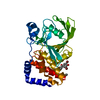
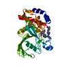
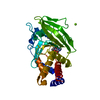
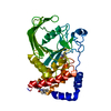
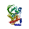
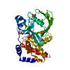
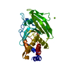
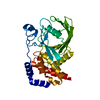
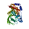
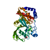
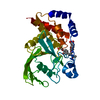
| Entry | Database: PDB / ID: 1ony | ||||||
|---|---|---|---|---|---|---|---|
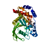
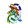
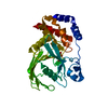
| Title | Oxalyl-Aryl-Amino Benzoic Acid inhibitors of PTP1B, compound 17 | ||||||
 Components Components | Protein-tyrosine phosphatase, non-receptor type 1 | ||||||
 Keywords Keywords | HYDROLASE / Protein Tyrosine Phosphatase / oxalyl-aryl-amino benzoic acid inhibitor | ||||||
| Function / homology |  Function and homology information Function and homology informationPTK6 Down-Regulation / regulation of hepatocyte growth factor receptor signaling pathway / positive regulation of receptor catabolic process / insulin receptor recycling / negative regulation of vascular endothelial growth factor receptor signaling pathway / regulation of intracellular protein transport / negative regulation of MAP kinase activity / mitochondrial crista / IRE1-mediated unfolded protein response / platelet-derived growth factor receptor-beta signaling pathway ...PTK6 Down-Regulation / regulation of hepatocyte growth factor receptor signaling pathway / positive regulation of receptor catabolic process / insulin receptor recycling / negative regulation of vascular endothelial growth factor receptor signaling pathway / regulation of intracellular protein transport / negative regulation of MAP kinase activity / mitochondrial crista / IRE1-mediated unfolded protein response / platelet-derived growth factor receptor-beta signaling pathway / sorting endosome / positive regulation of IRE1-mediated unfolded protein response / cytoplasmic side of endoplasmic reticulum membrane / negative regulation of PERK-mediated unfolded protein response / regulation of type I interferon-mediated signaling pathway / negative regulation of vascular associated smooth muscle cell migration / vascular endothelial cell response to oscillatory fluid shear stress / peptidyl-tyrosine dephosphorylation / positive regulation of systemic arterial blood pressure / non-membrane spanning protein tyrosine phosphatase activity / regulation of endocytosis / Regulation of IFNA/IFNB signaling / cellular response to angiotensin / regulation of proteolysis / growth hormone receptor signaling pathway via JAK-STAT / negative regulation of cell-substrate adhesion / cellular response to unfolded protein / regulation of postsynapse assembly / positive regulation of endothelial cell apoptotic process / regulation of signal transduction / negative regulation of signal transduction / Regulation of IFNG signaling / Growth hormone receptor signaling / negative regulation of endoplasmic reticulum stress-induced intrinsic apoptotic signaling pathway / positive regulation of heart rate / ephrin receptor binding / Insulin receptor recycling / MECP2 regulates neuronal receptors and channels / cellular response to platelet-derived growth factor stimulus / Integrin signaling / endoplasmic reticulum unfolded protein response / phosphoprotein phosphatase activity / protein-tyrosine-phosphatase / cellular response to fibroblast growth factor stimulus / cellular response to nitric oxide / negative regulation of insulin receptor signaling pathway / protein tyrosine phosphatase activity / positive regulation of cardiac muscle cell apoptotic process / protein phosphatase 2A binding / Turbulent (oscillatory, disturbed) flow shear stress activates signaling by PIEZO1 and integrins in endothelial cells / endosome lumen / negative regulation of phosphatidylinositol 3-kinase/protein kinase B signal transduction / insulin receptor binding / cellular response to nerve growth factor stimulus / response to nutrient levels / negative regulation of ERK1 and ERK2 cascade / Negative regulation of MET activity / receptor tyrosine kinase binding / positive regulation of JNK cascade / insulin receptor signaling pathway / negative regulation of neuron projection development / actin cytoskeleton organization / cellular response to hypoxia / early endosome / postsynapse / cadherin binding / mitochondrial matrix / negative regulation of cell population proliferation / protein kinase binding / glutamatergic synapse / enzyme binding / endoplasmic reticulum / protein-containing complex / RNA binding / zinc ion binding / cytoplasm / cytosol Similarity search - Function | ||||||
| Biological species |  Homo sapiens (human) Homo sapiens (human) | ||||||
| Method |  X-RAY DIFFRACTION / MOLECULAR REPLACEMENT / Resolution: 2.15 Å X-RAY DIFFRACTION / MOLECULAR REPLACEMENT / Resolution: 2.15 Å | ||||||
 Authors Authors | Liu, G. / Szczepankiewicz, B.G. / Pei, Z. / Janowich, D.A. / Xin, Z. / Hadjuk, P.J. / Abad-Zapatero, C. / Liang, H. / Hutchins, C.W. / Fesik, S.W. ...Liu, G. / Szczepankiewicz, B.G. / Pei, Z. / Janowich, D.A. / Xin, Z. / Hadjuk, P.J. / Abad-Zapatero, C. / Liang, H. / Hutchins, C.W. / Fesik, S.W. / Ballaron, S.J. / Stashko, M.A. / Lubben, T. / Mika, A.K. / Zinker, B.A. / Trevillyan, J.M. / Jirousek, M.R. | ||||||
 Citation Citation | Journal: J.Med.Chem. / Year: 2003 Title: Discovery and Structure-Activity Relationship of Oxalylarylaminobenzoic Acids as Inhibitors of Protein Tyrosine Phosphatase 1B Authors: Liu, G. / Szczepankiewicz, B.G. / Pei, Z. / Janowich, D.A. / Xin, Z. / Hadjuk, P.J. / Abad-Zapatero, C. / Liang, H. / Hutchins, C.W. / Fesik, S.W. / Ballaron, S.J. / Stashko, M.A. / Lubben, ...Authors: Liu, G. / Szczepankiewicz, B.G. / Pei, Z. / Janowich, D.A. / Xin, Z. / Hadjuk, P.J. / Abad-Zapatero, C. / Liang, H. / Hutchins, C.W. / Fesik, S.W. / Ballaron, S.J. / Stashko, M.A. / Lubben, T. / Mika, A.K. / Zinker, B.A. / Trevillyan, J.M. / Jirousek, M.R. #1: Journal: BIOORG.MED.CHEM.LETT. / Year: 2003Title: Potent, Selective Inhibitors of Protein Tyrosine Phosphatase 1B Authors: Xin, Z. / Oost, T.K. / Abad-Zapatero, C. / Hajduk, P.J. / Pei, Z. / Szczepankiewicz, B.G. / Hutchins, C.W. / Ballaron, S.J. / Stashko, M.A. / Lubben, T. / Trevillyan, J.M. / Jirousek, M.R. / Liu, G. #2: Journal: J.Am.Chem.Soc. / Year: 2003Title: Potent, Selective Protein Tyrosine Phosphatase 1B Inhibitor Using a Linked-Fragment Strategy Authors: Szczepankiewicz, B.G. / Liu, G. / Hajduk, P.J. / Abad-Zapatero, C. / Pei, Z. / Xin, Z. / Lubben, T. / Trevillyan, J.M. / Stashko, M.A. / Ballaron, S.J. / Liang, H. / Huang, F. / Hutchins, C. ...Authors: Szczepankiewicz, B.G. / Liu, G. / Hajduk, P.J. / Abad-Zapatero, C. / Pei, Z. / Xin, Z. / Lubben, T. / Trevillyan, J.M. / Stashko, M.A. / Ballaron, S.J. / Liang, H. / Huang, F. / Hutchins, C.W. / Fesik, S.W. / Jirousek, M.R. | ||||||
| History |
|
- Structure visualization
| Structure viewer | Molecule: MolmilJmol/JSmol |
|---|
- Downloads & links
-Download
| PDBx/mmCIF format | 1ony.cif.gz | 79.5 KB | Display | PDBx/mmCIF format |
|---|---|---|---|---|
| PDB format | pdb1ony.ent.gz | 58.4 KB | Display | PDB format |
| PDBx/mmJSON format | 1ony.json.gz | Tree view | PDBx/mmJSON format | |
| Others |  Other downloads Other downloads |
-Validation report
| Arichive directory | https://data.pdbj.org/pub/pdb/validation_reports/on/1onyftp://data.pdbj.org/pub/pdb/validation_reports/on/1ony | HTTPS FTP |
|---|
-Related structure data
| Related structure data |  1onzC 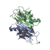 1tyrS C: citing same article ( S: Starting model for refinement |
|---|---|
| Similar structure data |


-Links
 PDBj
PDBj


- Assembly
| Deposited unit | 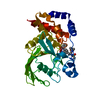
| ||||||||
|---|---|---|---|---|---|---|---|---|---|
| 1 |
| ||||||||
| Unit cell |
|
-Components
| #1: Protein | Mass: 37365.637 Da / Num. of mol.: 1 / Fragment: PTP1B Catalytic domain Source method: isolated from a genetically manipulated source Source: (gene. exp.) Homo sapiens (human) / Gene: PTPN1 OR PTP1B / Plasmid: pT7-7 / Production host:  |
|---|---|
| #2: Chemical | ChemComp-588 /   Mass: 588.629 Da / Num. of mol.: 1 / Source method: obtained synthetically / Formula: C27H32N4O9S Mass: 588.629 Da / Num. of mol.: 1 / Source method: obtained synthetically / Formula: C27H32N4O9S |
| #3: Water | ChemComp-HOH /  Mass: 18.015 Da / Num. of mol.: 312 / Source method: isolated from a natural source / Formula: H2O Mass: 18.015 Da / Num. of mol.: 312 / Source method: isolated from a natural source / Formula: H2O |
-Experimental details
-Experiment
| Experiment | Method: X-RAY DIFFRACTION / Number of used crystals: 1 |
|---|
- Sample preparation
| Crystal | Density Matthews: 3.22 Å3/Da / Density % sol: 61.83 % | ||||||||||||||||||||||||||||||||||||||||||||||||||||||||||||||||||||||||||||||
|---|---|---|---|---|---|---|---|---|---|---|---|---|---|---|---|---|---|---|---|---|---|---|---|---|---|---|---|---|---|---|---|---|---|---|---|---|---|---|---|---|---|---|---|---|---|---|---|---|---|---|---|---|---|---|---|---|---|---|---|---|---|---|---|---|---|---|---|---|---|---|---|---|---|---|---|---|---|---|---|
| Crystal grow | Temperature: 277 K / Method: vapor diffusion, hanging drop / pH: 7.1 Details: Precipitation buffer: 100 mM Hepes, 0.2 M Magnesium Acetate, 14% PEG8000, pH 7.1, VAPOR DIFFUSION, HANGING DROP, temperature 277K | ||||||||||||||||||||||||||||||||||||||||||||||||||||||||||||||||||||||||||||||
| Crystal grow | *PLUS Temperature: 4 ℃ / pH: 7.5 Details: Puius, Y.A., (1997) Proc.Natl.Acad.Sci.USA, 94, 13420. | ||||||||||||||||||||||||||||||||||||||||||||||||||||||||||||||||||||||||||||||
| Components of the solutions | *PLUS
|
-Data collection
| Diffraction | Mean temperature: 100 K |
|---|---|
| Diffraction source | Source: ROTATING ANODE / Type: RIGAKU RU300 / Wavelength: 1.5418 Å |
| Detector | Type: MARRESEARCH / Detector: CCD / Date: Aug 1, 2000 / Details: mirrors |
| Radiation | Protocol: SINGLE WAVELENGTH / Monochromatic (M) / Laue (L): M / Scattering type: x-ray |
| Radiation wavelength | Wavelength: 1.5418 Å / Relative weight: 1 |
| Reflection | Resolution: 2→50 Å / Num. all: 32536 / Num. obs: 30332 / % possible obs: 81.3 % / Observed criterion σ(F): 0 / Observed criterion σ(I): 1 / Redundancy: 4.8 % / Biso Wilson estimate: 33.3 Å2 / Rmerge(I) obs: 0.065 / Rsym value: 0.065 / Net I/σ(I): 16.9 |
| Reflection shell | Resolution: 2→2.07 Å / Redundancy: 4.1 % / Rmerge(I) obs: 0.571 / Mean I/σ(I) obs: 2.1 / Num. unique all: 3152 / Rsym value: 0.571 / % possible all: 96.5 |
- Processing
| Software |
| ||||||||||||||||||||||||||||||||||||
|---|---|---|---|---|---|---|---|---|---|---|---|---|---|---|---|---|---|---|---|---|---|---|---|---|---|---|---|---|---|---|---|---|---|---|---|---|---|
| Refinement | Method to determine structure: MOLECULAR REPLACEMENT Starting model: PDB entry 1TYR and initial internal refinement Resolution: 2.15→17.44 Å / Rfactor Rfree error: 0.005 / Isotropic thermal model: RESTRAINED / Cross valid method: THROUGHOUT / σ(F): 2 / σ(I): 0 / Stereochemistry target values: Engh & Huber Details: Residue CYS215, listed in remark 500, corresponds to the active site CYS which is known to be in a strained conformation in this class of enzymes. Strained conformation of Gly259 is caused ...Details: Residue CYS215, listed in remark 500, corresponds to the active site CYS which is known to be in a strained conformation in this class of enzymes. Strained conformation of Gly259 is caused by the presence of the inhibitor in the active site and in the vicinity of Gln262.
| ||||||||||||||||||||||||||||||||||||
| Solvent computation | Solvent model: FLAT MODEL / Bsol: 73.0446 Å2 / ksol: 0.388378 e/Å3 | ||||||||||||||||||||||||||||||||||||
| Displacement parameters | Biso mean: 36.2 Å2
| ||||||||||||||||||||||||||||||||||||
| Refine analyze |
| ||||||||||||||||||||||||||||||||||||
| Refinement step | Cycle: LAST / Resolution: 2.15→17.44 Å
| ||||||||||||||||||||||||||||||||||||
| Refine LS restraints |
| ||||||||||||||||||||||||||||||||||||
| LS refinement shell | Resolution: 2.15→2.23 Å / Rfactor Rfree error: 0.017 / Total num. of bins used: 10
| ||||||||||||||||||||||||||||||||||||
| Xplor file |
| ||||||||||||||||||||||||||||||||||||
| Refinement | *PLUS Rfactor Rfree: 0.22 | ||||||||||||||||||||||||||||||||||||
| Solvent computation | *PLUS | ||||||||||||||||||||||||||||||||||||
| Displacement parameters | *PLUS | ||||||||||||||||||||||||||||||||||||
| Refine LS restraints | *PLUS
|
