 Movie
Movie Controller

[English] 日本語
 Yorodumi
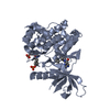
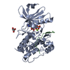
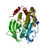
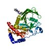
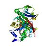
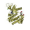
- PDB-1lqf: Structure of PTP1b in Complex with a Peptidic Bisphosphonate Inhibitor -
+ Open data

- Basic information
| Entry | Database: PDB / ID: 1lqf | ||||||
|---|---|---|---|---|---|---|---|
| Title | Structure of PTP1b in Complex with a Peptidic Bisphosphonate Inhibitor | ||||||
 Components Components | protein-tyrosine phosphatase, non-receptor type 1 | ||||||
 Keywords Keywords | HYDROLASE / Phosphatase / phosphonates / diabetes / inhibitor | ||||||
| Function / homology |  Function and homology information Function and homology informationPTK6 Down-Regulation / regulation of hepatocyte growth factor receptor signaling pathway / positive regulation of receptor catabolic process / insulin receptor recycling / negative regulation of vascular endothelial growth factor receptor signaling pathway / regulation of intracellular protein transport / negative regulation of MAP kinase activity / mitochondrial crista / IRE1-mediated unfolded protein response / platelet-derived growth factor receptor-beta signaling pathway ...PTK6 Down-Regulation / regulation of hepatocyte growth factor receptor signaling pathway / positive regulation of receptor catabolic process / insulin receptor recycling / negative regulation of vascular endothelial growth factor receptor signaling pathway / regulation of intracellular protein transport / negative regulation of MAP kinase activity / mitochondrial crista / IRE1-mediated unfolded protein response / platelet-derived growth factor receptor-beta signaling pathway / sorting endosome / positive regulation of IRE1-mediated unfolded protein response / cytoplasmic side of endoplasmic reticulum membrane / negative regulation of PERK-mediated unfolded protein response / regulation of type I interferon-mediated signaling pathway / negative regulation of vascular associated smooth muscle cell migration / vascular endothelial cell response to oscillatory fluid shear stress / peptidyl-tyrosine dephosphorylation / positive regulation of systemic arterial blood pressure / non-membrane spanning protein tyrosine phosphatase activity / regulation of endocytosis / Regulation of IFNA/IFNB signaling / cellular response to angiotensin / regulation of proteolysis / growth hormone receptor signaling pathway via JAK-STAT / negative regulation of cell-substrate adhesion / cellular response to unfolded protein / regulation of postsynapse assembly / positive regulation of endothelial cell apoptotic process / regulation of signal transduction / negative regulation of signal transduction / Regulation of IFNG signaling / Growth hormone receptor signaling / negative regulation of endoplasmic reticulum stress-induced intrinsic apoptotic signaling pathway / positive regulation of heart rate / ephrin receptor binding / Insulin receptor recycling / MECP2 regulates neuronal receptors and channels / cellular response to platelet-derived growth factor stimulus / Integrin signaling / endoplasmic reticulum unfolded protein response / phosphoprotein phosphatase activity / protein-tyrosine-phosphatase / cellular response to fibroblast growth factor stimulus / cellular response to nitric oxide / negative regulation of insulin receptor signaling pathway / protein tyrosine phosphatase activity / positive regulation of cardiac muscle cell apoptotic process / protein phosphatase 2A binding / Turbulent (oscillatory, disturbed) flow shear stress activates signaling by PIEZO1 and integrins in endothelial cells / endosome lumen / negative regulation of phosphatidylinositol 3-kinase/protein kinase B signal transduction / insulin receptor binding / cellular response to nerve growth factor stimulus / response to nutrient levels / negative regulation of ERK1 and ERK2 cascade / Negative regulation of MET activity / receptor tyrosine kinase binding / positive regulation of JNK cascade / insulin receptor signaling pathway / negative regulation of neuron projection development / actin cytoskeleton organization / cellular response to hypoxia / early endosome / postsynapse / cadherin binding / mitochondrial matrix / negative regulation of cell population proliferation / protein kinase binding / glutamatergic synapse / enzyme binding / endoplasmic reticulum / protein-containing complex / RNA binding / zinc ion binding / cytoplasm / cytosol Similarity search - Function | ||||||
| Biological species |  Homo sapiens (human) Homo sapiens (human) | ||||||
| Method |  X-RAY DIFFRACTION / SYNCHROTRON / MOLECULAR REPLACEMENT / Resolution: 2.5 Å X-RAY DIFFRACTION / SYNCHROTRON / MOLECULAR REPLACEMENT / Resolution: 2.5 Å | ||||||
 Authors Authors | Asante-Appiah, E. / Patel, S. / Dufresne, C. / Scapin, G. | ||||||
 Citation Citation | Journal: Biochemistry / Year: 2002 Title: The structure of PTP-1B in complex with a peptide inhibitor reveals an alternative binding mode for bisphosphonates. Authors: Asante-Appiah, E. / Patel, S. / Dufresne, C. / Roy, P. / Wang, Q. / Patel, V. / Friesen, R.W. / Ramachandran, C. / Becker, J.W. / Leblanc, Y. / Kennedy, B.P. / Scapin, G. #1: Journal: J.Biol.Chem. / Year: 2001Title: The YRD motif is a major determinant of substrate and inhibitor specificity in T-cell protein-tyrosine phosphatase Authors: Asante-Appiah, E. / Ball, K. / Bateman, K. / Skorey, K. / Friesen, R. / Desponts, C. / Payette, P. / Bayly, C. / Zamboni, R. / Scapin, G. / Ramachandran, C. / Kennedy, B.P. | ||||||
| History |
|
- Structure visualization
| Structure viewer | Molecule: MolmilJmol/JSmol |
|---|
- Downloads & links
-Download
| PDBx/mmCIF format | 1lqf.cif.gz | 255.2 KB | Display | PDBx/mmCIF format |
|---|---|---|---|---|
| PDB format | pdb1lqf.ent.gz | 206.6 KB | Display | PDB format |
| PDBx/mmJSON format | 1lqf.json.gz | Tree view | PDBx/mmJSON format | |
| Others |  Other downloads Other downloads |
-Validation report
| Arichive directory | https://data.pdbj.org/pub/pdb/validation_reports/lq/1lqfftp://data.pdbj.org/pub/pdb/validation_reports/lq/1lqf | HTTPS FTP |
|---|
-Related structure data
| Related structure data |  1ptyS S: Starting model for refinement |
|---|---|
| Similar structure data |


-Links
 PDBj
PDBj


- Assembly
| Deposited unit | 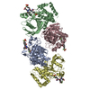
| |||||||||
|---|---|---|---|---|---|---|---|---|---|---|
| 1 | 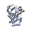
| |||||||||
| 2 | 
| |||||||||
| 3 | 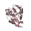
| |||||||||
| 4 | 
| |||||||||
| Unit cell |
| |||||||||
| Components on special symmetry positions |
|
-Components
| #1: Protein | Mass: 34451.367 Da / Num. of mol.: 4 / Fragment: catalytic domain (residues 1-283) Source method: isolated from a genetically manipulated source Source: (gene. exp.) Homo sapiens (human) / Species (production host): Escherichia coli / Production host:  #2: Chemical | ChemComp-BGD / 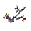  Mass: 804.573 Da / Num. of mol.: 4 / Source method: obtained synthetically / Formula: C32H34F4N4O12P2 Mass: 804.573 Da / Num. of mol.: 4 / Source method: obtained synthetically / Formula: C32H34F4N4O12P2#3: Water | ChemComp-HOH / |  Mass: 18.015 Da / Num. of mol.: 528 / Source method: isolated from a natural source / Formula: H2O Mass: 18.015 Da / Num. of mol.: 528 / Source method: isolated from a natural source / Formula: H2O |
|---|
-Experimental details
-Experiment
| Experiment | Method: X-RAY DIFFRACTION / Number of used crystals: 1 |
|---|
- Sample preparation
| Crystal | Density Matthews: 3.7 Å3/Da / Density % sol: 73 % | |||||||||||||||||||||||||||||||||||||||||||||||||||||||||||||||
|---|---|---|---|---|---|---|---|---|---|---|---|---|---|---|---|---|---|---|---|---|---|---|---|---|---|---|---|---|---|---|---|---|---|---|---|---|---|---|---|---|---|---|---|---|---|---|---|---|---|---|---|---|---|---|---|---|---|---|---|---|---|---|---|---|
| Crystal grow | Temperature: 284 K / Method: vapor diffusion, sitting drop / pH: 5.9 Details: Peg 4000, propanol, citrate, pH 5.9, VAPOR DIFFUSION, SITTING DROP, temperature 284K | |||||||||||||||||||||||||||||||||||||||||||||||||||||||||||||||
| Crystal grow | *PLUS Temperature: 4 ℃ / pH: 7 | |||||||||||||||||||||||||||||||||||||||||||||||||||||||||||||||
| Components of the solutions | *PLUS
|
-Data collection
| Diffraction | Mean temperature: 100 K |
|---|---|
| Diffraction source | Source: SYNCHROTRON / Site: APS  / Beamline: 17-ID / Wavelength: 1 Å / Beamline: 17-ID / Wavelength: 1 Å |
| Detector | Type: MARRESEARCH / Detector: CCD / Date: Dec 9, 1998 |
| Radiation | Protocol: SINGLE WAVELENGTH / Monochromatic (M) / Laue (L): M / Scattering type: x-ray |
| Radiation wavelength | Wavelength: 1 Å / Relative weight: 1 |
| Reflection | Resolution: 2.5→29 Å / Num. all: 63052 / Num. obs: 61097 / % possible obs: 96.9 % / Observed criterion σ(F): 0 / Observed criterion σ(I): 0 / Redundancy: 2.8 % / Biso Wilson estimate: 38.9 Å2 / Rsym value: 0.121 / Net I/σ(I): 6 |
| Reflection shell | Resolution: 2.5→2.65 Å / Redundancy: 1.9 % / Mean I/σ(I) obs: 1.5 / Num. unique all: 9039 / Rsym value: 0.38 / % possible all: 86.4 |
| Reflection | *PLUS Lowest resolution: 30 Å / Num. measured all: 176274 / Rmerge(I) obs: 0.121 |
| Reflection shell | *PLUS % possible obs: 86.4 % / Num. unique obs: 9039 / Num. measured obs: 17193 / Rmerge(I) obs: 0.38 |
- Processing
| Software |
| |||||||||||||||||||||||||
|---|---|---|---|---|---|---|---|---|---|---|---|---|---|---|---|---|---|---|---|---|---|---|---|---|---|---|
| Refinement | Method to determine structure: MOLECULAR REPLACEMENT Starting model: 1PTY Resolution: 2.5→29 Å / Isotropic thermal model: Anisotropic / Cross valid method: THROUGHOUT / σ(F): 0 / Stereochemistry target values: Engh & Huber Details: A 4% twinning was detected using both Truncate and CNS and the refinement was carried out using the CNS twinning procedures, with twinning operator h, -k, -l.
| |||||||||||||||||||||||||
| Displacement parameters | Biso mean: 42.1 Å2
| |||||||||||||||||||||||||
| Refinement step | Cycle: LAST / Resolution: 2.5→29 Å
| |||||||||||||||||||||||||
| Refine LS restraints |
| |||||||||||||||||||||||||
| LS refinement shell | Resolution: 2.5→2.68 Å / Total num. of bins used: 10
| |||||||||||||||||||||||||
| Refinement | *PLUS Lowest resolution: 30 Å / Num. reflection obs: 61052 / % reflection Rfree: 5 % / Rfactor obs: 0.231 / Rfactor Rfree: 0.286 / Rfactor Rwork: 0.228 | |||||||||||||||||||||||||
| Solvent computation | *PLUS | |||||||||||||||||||||||||
| Displacement parameters | *PLUS | |||||||||||||||||||||||||
| Refine LS restraints | *PLUS
| |||||||||||||||||||||||||
| LS refinement shell | *PLUS Rfactor Rfree: 0.438 / Rfactor Rwork: 0.366 |
