 Movie
Movie Controller

[English] 日本語
 Yorodumi
- PDB-3u1s: Crystal structure of human Fab PGT145, a broadly reactive and pot... -
+ Open data

- Basic information
| Entry | Database: PDB / ID: 3u1s | ||||||
|---|---|---|---|---|---|---|---|
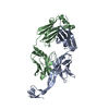
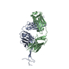
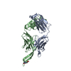
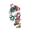
| Title | Crystal structure of human Fab PGT145, a broadly reactive and potent HIV-1 neutralizing antibody | ||||||
 Components Components |
| ||||||
 Keywords Keywords | IMMUNE SYSTEM / IgG / broadly neutralizing antibody / HIV-1 gp120 | ||||||
| Function / homology | Immunoglobulins / Immunoglobulin-like / Sandwich / Mainly Beta Function and homology information Function and homology information | ||||||
| Biological species |  Homo sapiens (human) Homo sapiens (human) | ||||||
| Method |  X-RAY DIFFRACTION / SYNCHROTRON / MOLECULAR REPLACEMENT / Resolution: 2.3 Å X-RAY DIFFRACTION / SYNCHROTRON / MOLECULAR REPLACEMENT / Resolution: 2.3 Å | ||||||
 Authors Authors | Julien, J.-P. / Diwanji, D. / Burton, D.R. / Wilson, I.A. | ||||||
 Citation Citation | Journal: Nature / Year: 2011 Title: Structure of HIV-1 gp120 V1/V2 domain with broadly neutralizing antibody PG9. Authors: McLellan, J.S. / Pancera, M. / Carrico, C. / Gorman, J. / Julien, J.P. / Khayat, R. / Louder, R. / Pejchal, R. / Sastry, M. / Dai, K. / O'Dell, S. / Patel, N. / Shahzad-Ul-Hussan, S. / Yang, ...Authors: McLellan, J.S. / Pancera, M. / Carrico, C. / Gorman, J. / Julien, J.P. / Khayat, R. / Louder, R. / Pejchal, R. / Sastry, M. / Dai, K. / O'Dell, S. / Patel, N. / Shahzad-Ul-Hussan, S. / Yang, Y. / Zhang, B. / Zhou, T. / Zhu, J. / Boyington, J.C. / Chuang, G.Y. / Diwanji, D. / Georgiev, I. / Do Kwon, Y. / Lee, D. / Louder, M.K. / Moquin, S. / Schmidt, S.D. / Yang, Z.Y. / Bonsignori, M. / Crump, J.A. / Kapiga, S.H. / Sam, N.E. / Haynes, B.F. / Burton, D.R. / Koff, W.C. / Walker, L.M. / Phogat, S. / Wyatt, R. / Orwenyo, J. / Wang, L.X. / Arthos, J. / Bewley, C.A. / Mascola, J.R. / Nabel, G.J. / Schief, W.R. / Ward, A.B. / Wilson, I.A. / Kwong, P.D. | ||||||
| History |
|
- Structure visualization
| Structure viewer | Molecule: MolmilJmol/JSmol |
|---|
- Downloads & links
-Download
| PDBx/mmCIF format | 3u1s.cif.gz | 197.8 KB | Display | PDBx/mmCIF format |
|---|---|---|---|---|
| PDB format | pdb3u1s.ent.gz | 156.7 KB | Display | PDB format |
| PDBx/mmJSON format | 3u1s.json.gz | Tree view | PDBx/mmJSON format | |
| Others |  Other downloads Other downloads |
-Validation report
| Arichive directory | https://data.pdbj.org/pub/pdb/validation_reports/u1/3u1sftp://data.pdbj.org/pub/pdb/validation_reports/u1/3u1s | HTTPS FTP |
|---|
-Related structure data
| Related structure data |  3tclC  3u2sC  3u36C  3u46C  3u4bC  3u4eC  3mugS S: Starting model for refinement C: citing same article ( |
|---|---|
| Similar structure data |
-Links
 PDBj
PDBj


- Assembly
| Deposited unit | 
| ||||||||
|---|---|---|---|---|---|---|---|---|---|
| 1 |
| ||||||||
| Unit cell |
|
-Components
| #1: Antibody | Mass: 26050.344 Da / Num. of mol.: 1 / Fragment: fragment antigen binding Source method: isolated from a genetically manipulated source Source: (gene. exp.) Homo sapiens (human) / Cell line (production host): HEK 293T / Production host: Homo sapiens (human) | ||||||
|---|---|---|---|---|---|---|---|
| #2: Antibody | Mass: 28953.285 Da / Num. of mol.: 1 / Fragment: fragment antigen binding Source method: isolated from a genetically manipulated source Source: (gene. exp.) Homo sapiens (human) / Cell line (production host): HEK 293T / Production host: Homo sapiens (human) | ||||||
| #3: Chemical | ChemComp-SO4 /   Mass: 96.063 Da / Num. of mol.: 6 / Source method: obtained synthetically / Formula: SO4 Mass: 96.063 Da / Num. of mol.: 6 / Source method: obtained synthetically / Formula: SO4#4: Chemical | ChemComp-GOL / |   Mass: 92.094 Da / Num. of mol.: 1 / Source method: obtained synthetically / Formula: C3H8O3 Mass: 92.094 Da / Num. of mol.: 1 / Source method: obtained synthetically / Formula: C3H8O3#5: Water | ChemComp-HOH / |  Mass: 18.015 Da / Num. of mol.: 313 / Source method: isolated from a natural source / Formula: H2O Mass: 18.015 Da / Num. of mol.: 313 / Source method: isolated from a natural source / Formula: H2OHas protein modification | Y | |
-Experimental details
-Experiment
| Experiment | Method: X-RAY DIFFRACTION / Number of used crystals: 1 |
|---|
- Sample preparation
| Crystal | Density Matthews: 3.24 Å3/Da / Density % sol: 62.05 % |
|---|---|
| Crystal grow | Temperature: 293 K / Method: vapor diffusion, sitting drop / pH: 7.5 Details: 0.1 M HEPES, pH 7.5, 2 M ammonium sulfate and 20% PEG 400, VAPOR DIFFUSION, SITTING DROP, temperature 293K |
-Data collection
| Diffraction | Mean temperature: 100 K |
|---|---|
| Diffraction source | Source: SYNCHROTRON / Site: APS  / Beamline: 23-ID-D / Wavelength: 1.03322 Å / Beamline: 23-ID-D / Wavelength: 1.03322 Å |
| Detector | Type: MARMOSAIC 300 mm CCD / Detector: CCD / Date: Aug 4, 2011 |
| Radiation | Monochromator: Double crystal cryo-cooled / Protocol: SINGLE WAVELENGTH / Monochromatic (M) / Laue (L): M / Scattering type: x-ray |
| Radiation wavelength | Wavelength: 1.03322 Å / Relative weight: 1 |
| Reflection | Resolution: 2.3→50 Å / Num. all: 32853 / Num. obs: 32493 / % possible obs: 99.2 % / Redundancy: 8.4 % / Rmerge(I) obs: 0.087 / Net I/σ(I): 19.1 |
| Reflection shell | Resolution: 2.3→2.4 Å / Redundancy: 8.6 % / Rmerge(I) obs: 0.472 / Mean I/σ(I) obs: 4.1 / % possible all: 99.9 |
- Processing
| Software |
| ||||||||||||||||||||||||||||||||||||||||||||||||||||||||||||||||||||||||||||||||||||||||||||||||||||||||||||||||||||||||||||||||||||||||||||||||||||||||||||||||||||||||||
|---|---|---|---|---|---|---|---|---|---|---|---|---|---|---|---|---|---|---|---|---|---|---|---|---|---|---|---|---|---|---|---|---|---|---|---|---|---|---|---|---|---|---|---|---|---|---|---|---|---|---|---|---|---|---|---|---|---|---|---|---|---|---|---|---|---|---|---|---|---|---|---|---|---|---|---|---|---|---|---|---|---|---|---|---|---|---|---|---|---|---|---|---|---|---|---|---|---|---|---|---|---|---|---|---|---|---|---|---|---|---|---|---|---|---|---|---|---|---|---|---|---|---|---|---|---|---|---|---|---|---|---|---|---|---|---|---|---|---|---|---|---|---|---|---|---|---|---|---|---|---|---|---|---|---|---|---|---|---|---|---|---|---|---|---|---|---|---|---|---|---|---|
| Refinement | Method to determine structure: MOLECULAR REPLACEMENT Starting model: 3MUG Resolution: 2.3→50 Å / Cor.coef. Fo:Fc: 0.946 / Cor.coef. Fo:Fc free: 0.926 / SU B: 11.849 / SU ML: 0.129 / Cross valid method: THROUGHOUT / ESU R: 0.225 / ESU R Free: 0.188 / Stereochemistry target values: MAXIMUM LIKELIHOOD / Details: HYDROGENS HAVE BEEN ADDED IN THE RIDING POSITIONS
| ||||||||||||||||||||||||||||||||||||||||||||||||||||||||||||||||||||||||||||||||||||||||||||||||||||||||||||||||||||||||||||||||||||||||||||||||||||||||||||||||||||||||||
| Solvent computation | Ion probe radii: 0.8 Å / Shrinkage radii: 0.8 Å / VDW probe radii: 1.4 Å / Solvent model: MASK | ||||||||||||||||||||||||||||||||||||||||||||||||||||||||||||||||||||||||||||||||||||||||||||||||||||||||||||||||||||||||||||||||||||||||||||||||||||||||||||||||||||||||||
| Displacement parameters | Biso mean: 34.339 Å2
| ||||||||||||||||||||||||||||||||||||||||||||||||||||||||||||||||||||||||||||||||||||||||||||||||||||||||||||||||||||||||||||||||||||||||||||||||||||||||||||||||||||||||||
| Refinement step | Cycle: LAST / Resolution: 2.3→50 Å
| ||||||||||||||||||||||||||||||||||||||||||||||||||||||||||||||||||||||||||||||||||||||||||||||||||||||||||||||||||||||||||||||||||||||||||||||||||||||||||||||||||||||||||
| Refine LS restraints |
| ||||||||||||||||||||||||||||||||||||||||||||||||||||||||||||||||||||||||||||||||||||||||||||||||||||||||||||||||||||||||||||||||||||||||||||||||||||||||||||||||||||||||||
| LS refinement shell | Resolution: 2.3→2.36 Å / Total num. of bins used: 20
| ||||||||||||||||||||||||||||||||||||||||||||||||||||||||||||||||||||||||||||||||||||||||||||||||||||||||||||||||||||||||||||||||||||||||||||||||||||||||||||||||||||||||||
| Refinement TLS params. | Method: refined / Refine-ID: X-RAY DIFFRACTION
| ||||||||||||||||||||||||||||||||||||||||||||||||||||||||||||||||||||||||||||||||||||||||||||||||||||||||||||||||||||||||||||||||||||||||||||||||||||||||||||||||||||||||||
| Refinement TLS group |
|
