 Movie
Movie Controller

[English] 日本語
 Yorodumi
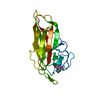
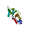
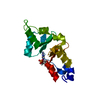
- PDB-3twu: Crystal structure of ARC4 from human Tankyrase 2 in complex with ... -
+ Open data

- Basic information
| Entry | Database: PDB / ID: 3twu | ||||||
|---|---|---|---|---|---|---|---|
| Title | Crystal structure of ARC4 from human Tankyrase 2 in complex with peptide from human MCL1 | ||||||
 Components Components |
| ||||||
 Keywords Keywords | SIGNALING PROTEIN/PEPTIDE / ankyrin repeat / protein-protein interaction / substrate recruitment / poly(ADP-ribosyl)ation / SIGNALING PROTEIN-PEPTIDE complex | ||||||
| Function / homology |  Function and homology information Function and homology informationpositive regulation of oxidative stress-induced neuron intrinsic apoptotic signaling pathway / XAV939 stabilizes AXIN / cell fate determination / positive regulation of telomere capping / cellular homeostasis / Bcl-2 family protein complex / mitochondrial fusion / NAD+ ADP-ribosyltransferase / protein auto-ADP-ribosylation / protein localization to chromosome, telomeric region ...positive regulation of oxidative stress-induced neuron intrinsic apoptotic signaling pathway / XAV939 stabilizes AXIN / cell fate determination / positive regulation of telomere capping / cellular homeostasis / Bcl-2 family protein complex / mitochondrial fusion / NAD+ ADP-ribosyltransferase / protein auto-ADP-ribosylation / protein localization to chromosome, telomeric region / negative regulation of telomere maintenance via telomere lengthening / NAD+-protein-aspartate ADP-ribosyltransferase activity / protein poly-ADP-ribosylation / NAD+-protein-glutamate ADP-ribosyltransferase activity / NAD+-protein mono-ADP-ribosyltransferase activity / pericentriolar material / Transferases; Glycosyltransferases; Pentosyltransferases / negative regulation of anoikis / BH3 domain binding / NAD+ poly-ADP-ribosyltransferase activity / transmembrane protein transporter activity / negative regulation of extrinsic apoptotic signaling pathway in absence of ligand / extrinsic apoptotic signaling pathway in absence of ligand / positive regulation of telomere maintenance via telomerase / nucleotidyltransferase activity / response to cytokine / release of cytochrome c from mitochondria / negative regulation of autophagy / TCF dependent signaling in response to WNT / Degradation of AXIN / intrinsic apoptotic signaling pathway in response to DNA damage / Wnt signaling pathway / Regulation of PTEN stability and activity / protein polyubiquitination / Signaling by ALK fusions and activated point mutants / nuclear envelope / positive regulation of canonical Wnt signaling pathway / positive regulation of neuron apoptotic process / channel activity / Interleukin-4 and Interleukin-13 signaling / regulation of apoptotic process / mitochondrial outer membrane / chromosome, telomeric region / Ub-specific processing proteases / positive regulation of apoptotic process / protein heterodimerization activity / Golgi membrane / DNA damage response / negative regulation of apoptotic process / perinuclear region of cytoplasm / enzyme binding / mitochondrion / nucleoplasm / membrane / metal ion binding / nucleus / cytoplasm / cytosol Similarity search - Function | ||||||
| Biological species |  Homo sapiens (human) Homo sapiens (human) | ||||||
| Method |  X-RAY DIFFRACTION / SYNCHROTRON / MOLECULAR REPLACEMENT / Resolution: 1.8 Å X-RAY DIFFRACTION / SYNCHROTRON / MOLECULAR REPLACEMENT / Resolution: 1.8 Å | ||||||
 Authors Authors | Guettler, S. / Sicheri, F. | ||||||
 Citation Citation | Journal: Cell(Cambridge,Mass.) / Year: 2011 Title: Structural basis and sequence rules for substrate recognition by tankyrase explain the basis for cherubism disease. Authors: Guettler, S. / Larose, J. / Petsalaki, E. / Gish, G. / Scotter, A. / Pawson, T. / Rottapel, R. / Sicheri, F. | ||||||
| History |
|
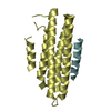
- Structure visualization
| Structure viewer | Molecule: MolmilJmol/JSmol |
|---|
- Downloads & links
-Download
| PDBx/mmCIF format | 3twu.cif.gz | 81.5 KB | Display | PDBx/mmCIF format |
|---|---|---|---|---|
| PDB format | pdb3twu.ent.gz | 61.6 KB | Display | PDB format |
| PDBx/mmJSON format | 3twu.json.gz | Tree view | PDBx/mmJSON format | |
| Others |  Other downloads Other downloads |
-Validation report
| Arichive directory | https://data.pdbj.org/pub/pdb/validation_reports/tw/3twuftp://data.pdbj.org/pub/pdb/validation_reports/tw/3twu | HTTPS FTP |
|---|
-Related structure data
| Related structure data |  3twqC  3twrC  3twsC  3twtC  3twvC  3twwC  3twxC C: citing same article ( |
|---|---|
| Similar structure data |


-Links
 PDBj
PDBj


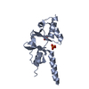
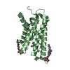
- Assembly
| Deposited unit | 
| ||||||||
|---|---|---|---|---|---|---|---|---|---|
| 1 |
| ||||||||
| Unit cell |
|
-Components
| #1: Protein | Mass: 17995.311 Da / Num. of mol.: 1 / Fragment: UNP residues 488-649 Source method: isolated from a genetically manipulated source Source: (gene. exp.) Homo sapiens (human) / Gene: PARP5B, TANK2, TNKL, TNKS2 / Plasmid: pETM-30-2 / Production host:  |
|---|---|
| #2: Protein/peptide | Mass: 1718.977 Da / Num. of mol.: 1 / Fragment: UNP residues 73-88 / Source method: obtained synthetically / Details: solid-state synthesized peptide / Source: (synth.) Homo sapiens (human) / References: UniProt: Q07820 |
| #3: Water | ChemComp-HOH /  Mass: 18.015 Da / Num. of mol.: 109 / Source method: isolated from a natural source / Formula: H2O Mass: 18.015 Da / Num. of mol.: 109 / Source method: isolated from a natural source / Formula: H2O |
| Has protein modification | Y |
-Experimental details
-Experiment
| Experiment | Method: X-RAY DIFFRACTION / Number of used crystals: 1 |
|---|
- Sample preparation
| Crystal | Density Matthews: 1.87 Å3/Da / Density % sol: 34.11 % |
|---|---|
| Crystal grow | Temperature: 293 K / Method: vapor diffusion, hanging drop / pH: 6.5 Details: 0.1 M HEPES-NaOH pH 6.5, 0.2 M NaOAc, 35% PEG 4000, VAPOR DIFFUSION, HANGING DROP, temperature 293K |
-Data collection
| Diffraction | Mean temperature: 100 K |
|---|---|
| Diffraction source | Source: SYNCHROTRON / Site: APS  / Beamline: 24-ID-E / Wavelength: 0.9792 Å / Beamline: 24-ID-E / Wavelength: 0.9792 Å |
| Detector | Type: ADSC QUANTUM 315 / Detector: CCD / Date: Mar 19, 2011 |
| Radiation | Monochromator: Si (220), Si (311) / Protocol: SINGLE WAVELENGTH / Monochromatic (M) / Laue (L): M / Scattering type: x-ray |
| Radiation wavelength | Wavelength: 0.9792 Å / Relative weight: 1 |
| Reflection | Resolution: 1.8→50 Å / Num. all: 14331 / Num. obs: 14331 / % possible obs: 97.2 % / Observed criterion σ(F): 0 / Observed criterion σ(I): 2.4 / Redundancy: 6.9 % / Rmerge(I) obs: 0.067 / Net I/σ(I): 23 |
| Reflection shell | Resolution: 1.8→1.86 Å / Redundancy: 5.8 % / Rmerge(I) obs: 0.469 / Mean I/σ(I) obs: 2.4 / % possible all: 91.9 |
- Processing
| Software |
| |||||||||||||||||||||||||||||||||||||||||||||||||||||||||||||||||||||||||||||||||||||||||||||||||||||||||||||||||||||||||||||||||||||||||||||||||||||||||||||||||||||||||||||||||||||||||||||||||||||||||||||||||||||||||||||||||||||||||||||||||||||||||||||||||||||||||||||||||||||||||||||||||||||||||||||||||||||||||||||||||||||
|---|---|---|---|---|---|---|---|---|---|---|---|---|---|---|---|---|---|---|---|---|---|---|---|---|---|---|---|---|---|---|---|---|---|---|---|---|---|---|---|---|---|---|---|---|---|---|---|---|---|---|---|---|---|---|---|---|---|---|---|---|---|---|---|---|---|---|---|---|---|---|---|---|---|---|---|---|---|---|---|---|---|---|---|---|---|---|---|---|---|---|---|---|---|---|---|---|---|---|---|---|---|---|---|---|---|---|---|---|---|---|---|---|---|---|---|---|---|---|---|---|---|---|---|---|---|---|---|---|---|---|---|---|---|---|---|---|---|---|---|---|---|---|---|---|---|---|---|---|---|---|---|---|---|---|---|---|---|---|---|---|---|---|---|---|---|---|---|---|---|---|---|---|---|---|---|---|---|---|---|---|---|---|---|---|---|---|---|---|---|---|---|---|---|---|---|---|---|---|---|---|---|---|---|---|---|---|---|---|---|---|---|---|---|---|---|---|---|---|---|---|---|---|---|---|---|---|---|---|---|---|---|---|---|---|---|---|---|---|---|---|---|---|---|---|---|---|---|---|---|---|---|---|---|---|---|---|---|---|---|---|---|---|---|---|---|---|---|---|---|---|---|---|---|---|---|---|---|---|---|---|---|---|---|---|---|---|---|---|---|---|---|---|---|---|---|---|---|---|---|---|---|---|---|---|---|---|---|---|---|---|---|---|---|---|---|---|---|---|---|---|---|---|---|---|---|---|
| Refinement | Method to determine structure: MOLECULAR REPLACEMENT / Resolution: 1.8→47.831 Å / SU ML: 0.43 / σ(F): 0.54 / Phase error: 18.65 / Stereochemistry target values: ML
| |||||||||||||||||||||||||||||||||||||||||||||||||||||||||||||||||||||||||||||||||||||||||||||||||||||||||||||||||||||||||||||||||||||||||||||||||||||||||||||||||||||||||||||||||||||||||||||||||||||||||||||||||||||||||||||||||||||||||||||||||||||||||||||||||||||||||||||||||||||||||||||||||||||||||||||||||||||||||||||||||||||
| Solvent computation | Shrinkage radii: 0.72 Å / VDW probe radii: 1 Å / Solvent model: FLAT BULK SOLVENT MODEL / Bsol: 58.39 Å2 / ksol: 0.388 e/Å3 | |||||||||||||||||||||||||||||||||||||||||||||||||||||||||||||||||||||||||||||||||||||||||||||||||||||||||||||||||||||||||||||||||||||||||||||||||||||||||||||||||||||||||||||||||||||||||||||||||||||||||||||||||||||||||||||||||||||||||||||||||||||||||||||||||||||||||||||||||||||||||||||||||||||||||||||||||||||||||||||||||||||
| Displacement parameters |
| |||||||||||||||||||||||||||||||||||||||||||||||||||||||||||||||||||||||||||||||||||||||||||||||||||||||||||||||||||||||||||||||||||||||||||||||||||||||||||||||||||||||||||||||||||||||||||||||||||||||||||||||||||||||||||||||||||||||||||||||||||||||||||||||||||||||||||||||||||||||||||||||||||||||||||||||||||||||||||||||||||||
| Refinement step | Cycle: LAST / Resolution: 1.8→47.831 Å
| |||||||||||||||||||||||||||||||||||||||||||||||||||||||||||||||||||||||||||||||||||||||||||||||||||||||||||||||||||||||||||||||||||||||||||||||||||||||||||||||||||||||||||||||||||||||||||||||||||||||||||||||||||||||||||||||||||||||||||||||||||||||||||||||||||||||||||||||||||||||||||||||||||||||||||||||||||||||||||||||||||||
| Refine LS restraints |
| |||||||||||||||||||||||||||||||||||||||||||||||||||||||||||||||||||||||||||||||||||||||||||||||||||||||||||||||||||||||||||||||||||||||||||||||||||||||||||||||||||||||||||||||||||||||||||||||||||||||||||||||||||||||||||||||||||||||||||||||||||||||||||||||||||||||||||||||||||||||||||||||||||||||||||||||||||||||||||||||||||||
| LS refinement shell |
| |||||||||||||||||||||||||||||||||||||||||||||||||||||||||||||||||||||||||||||||||||||||||||||||||||||||||||||||||||||||||||||||||||||||||||||||||||||||||||||||||||||||||||||||||||||||||||||||||||||||||||||||||||||||||||||||||||||||||||||||||||||||||||||||||||||||||||||||||||||||||||||||||||||||||||||||||||||||||||||||||||||
| Refinement TLS params. | Method: refined / Refine-ID: X-RAY DIFFRACTION
| |||||||||||||||||||||||||||||||||||||||||||||||||||||||||||||||||||||||||||||||||||||||||||||||||||||||||||||||||||||||||||||||||||||||||||||||||||||||||||||||||||||||||||||||||||||||||||||||||||||||||||||||||||||||||||||||||||||||||||||||||||||||||||||||||||||||||||||||||||||||||||||||||||||||||||||||||||||||||||||||||||||
| Refinement TLS group |
|
