 Movie
Movie Controller

[English] 日本語
 Yorodumi
- PDB-4xev: Fusion of Pyk2-FAT domain with Leupaxin LD1 motif, complexed with... -
+ Open data

- Basic information
| Entry | Database: PDB / ID: 4xev | ||||||
|---|---|---|---|---|---|---|---|
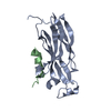
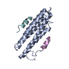
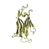
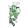
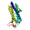
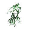
| Title | Fusion of Pyk2-FAT domain with Leupaxin LD1 motif, complexed with Leupaxin LD4 peptide | ||||||
 Components Components |
| ||||||
 Keywords Keywords | CELL ADHESION / 4-HELIX BUNDLE / FOCAL ADHESION / TYROSINE KINASE / LEUPAXIN | ||||||
| Function / homology |  Function and homology information Function and homology informationregulation of macrophage chemotaxis / neurotransmitter receptor regulator activity / positive regulation of B cell chemotaxis / negative regulation of B cell receptor signaling pathway / marginal zone B cell differentiation / regulation of ubiquitin-dependent protein catabolic process / endothelin receptor signaling pathway / negative regulation of myeloid cell differentiation / negative regulation of bone mineralization / regulation of postsynaptic density assembly ...regulation of macrophage chemotaxis / neurotransmitter receptor regulator activity / positive regulation of B cell chemotaxis / negative regulation of B cell receptor signaling pathway / marginal zone B cell differentiation / regulation of ubiquitin-dependent protein catabolic process / endothelin receptor signaling pathway / negative regulation of myeloid cell differentiation / negative regulation of bone mineralization / regulation of postsynaptic density assembly / activation of Janus kinase activity / cortical cytoskeleton organization / negative regulation of cell adhesion / signal complex assembly / sprouting angiogenesis / calcium/calmodulin-dependent protein kinase activity / apical dendrite / regulation of cell adhesion mediated by integrin / Interleukin-2 signaling / podosome / regulation of release of sequestered calcium ion into cytosol / peptidyl-tyrosine autophosphorylation / Signal regulatory protein family interactions / positive regulation of ubiquitin-dependent protein catabolic process / NMDA selective glutamate receptor complex / positive regulation of cell-matrix adhesion / positive regulation of actin filament polymerization / positive regulation of protein kinase activity / RHOU GTPase cycle / negative regulation of potassium ion transport / signaling receptor activator activity / bone resorption / vascular endothelial growth factor receptor signaling pathway / endothelial cell migration / regulation of cell adhesion / cellular defense response / postsynaptic density, intracellular component / cellular response to retinoic acid / substrate adhesion-dependent cell spreading / positive regulation of synaptic transmission, glutamatergic / peptidyl-tyrosine phosphorylation / transforming growth factor beta receptor signaling pathway / positive regulation of endothelial cell migration / ionotropic glutamate receptor signaling pathway / ionotropic glutamate receptor binding / integrin-mediated signaling pathway / positive regulation of excitatory postsynaptic potential / tumor necrosis factor-mediated signaling pathway / regulation of actin cytoskeleton organization / chemokine-mediated signaling pathway / non-specific protein-tyrosine kinase / transcription coregulator activity / non-membrane spanning protein tyrosine kinase activity / positive regulation of neuron projection development / regulation of synaptic plasticity / positive regulation of JNK cascade / VEGFA-VEGFR2 Pathway / epidermal growth factor receptor signaling pathway / long-term synaptic potentiation / positive regulation of angiogenesis / protein autophosphorylation / regulation of cell shape / lamellipodium / presynapse / growth cone / protein tyrosine kinase activity / protein-containing complex assembly / cell body / cell cortex / negative regulation of neuron apoptotic process / adaptive immune response / positive regulation of ERK1 and ERK2 cascade / positive regulation of phosphatidylinositol 3-kinase/protein kinase B signal transduction / cell surface receptor signaling pathway / cell adhesion / nuclear speck / postsynaptic density / positive regulation of cell migration / negative regulation of cell population proliferation / focal adhesion / neuronal cell body / apoptotic process / positive regulation of cell population proliferation / dendrite / negative regulation of apoptotic process / perinuclear region of cytoplasm / glutamatergic synapse / signal transduction / ATP binding / membrane / metal ion binding / nucleus / plasma membrane / cytosol / cytoplasm Similarity search - Function | ||||||
| Biological species |  Homo sapiens (human) Homo sapiens (human) | ||||||
| Method |  X-RAY DIFFRACTION / SYNCHROTRON / MOLECULAR REPLACEMENT / Resolution: 2.0073 Å X-RAY DIFFRACTION / SYNCHROTRON / MOLECULAR REPLACEMENT / Resolution: 2.0073 Å | ||||||
 Authors Authors | Miller, D.J. | ||||||
| Funding support |  United States, 1items United States, 1items
| ||||||
 Citation Citation | Journal: Biochemistry / Year: 2016 Title: Structural Basis for the Interaction between Pyk2-FAT Domain and Leupaxin LD Repeats. Authors: Vanarotti, M.S. / Finkelstein, D.B. / Guibao, C.D. / Nourse, A. / Miller, D.J. / Zheng, J.J. | ||||||
| History |
|
- Structure visualization
| Structure viewer | Molecule: MolmilJmol/JSmol |
|---|
- Downloads & links
-Download
| PDBx/mmCIF format | 4xev.cif.gz | 142.8 KB | Display | PDBx/mmCIF format |
|---|---|---|---|---|
| PDB format | pdb4xev.ent.gz | 111.9 KB | Display | PDB format |
| PDBx/mmJSON format | 4xev.json.gz | Tree view | PDBx/mmJSON format | |
| Others |  Other downloads Other downloads |
-Validation report
| Arichive directory | https://data.pdbj.org/pub/pdb/validation_reports/xe/4xevftp://data.pdbj.org/pub/pdb/validation_reports/xe/4xev | HTTPS FTP |
|---|
-Related structure data
| Related structure data | 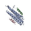 4xefC  4xekC  3gm3S C: citing same article ( S: Starting model for refinement |
|---|---|
| Similar structure data |


-Links
 PDBj
PDBj


- Assembly
| Deposited unit | 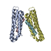
| ||||||||
|---|---|---|---|---|---|---|---|---|---|
| 1 | 
| ||||||||
| 2 | 
| ||||||||
| Unit cell |
|
-Components
| #1: Protein | Mass: 18034.441 Da / Num. of mol.: 2 / Mutation: C899S, C972A Source method: isolated from a genetically manipulated source Source: (gene. exp.) Homo sapiens (human) / Gene: PTK2B, FAK2, PYK2, RAFTK / Production host:  References: UniProt: Q14289, non-specific protein-tyrosine kinase #2: Protein/peptide | Mass: 2090.376 Da / Num. of mol.: 2 / Source method: obtained synthetically / Source: (synth.) Homo sapiens (human) / References: UniProt: O60711*PLUS#3: Water | ChemComp-HOH / |  Mass: 18.015 Da / Num. of mol.: 71 / Source method: isolated from a natural source / Formula: H2O Mass: 18.015 Da / Num. of mol.: 71 / Source method: isolated from a natural source / Formula: H2O |
|---|
-Experimental details
-Experiment
| Experiment | Method: X-RAY DIFFRACTION / Number of used crystals: 1 |
|---|
- Sample preparation
| Crystal | Density Matthews: 2.42 Å3/Da / Density % sol: 49.12 % |
|---|---|
| Crystal grow | Temperature: 291.2 K / Method: vapor diffusion, sitting drop / pH: 6.5 Details: The 4 ul drop contained 2 ul protein/LD4 peptide mixture (20 mM MES pH 6.2, 1 mM protein, and 2 mM peptide) and 2 ul well solution (100 mM MES pH 6.5 and 25 % PEG 3000). |
-Data collection
| Diffraction | Mean temperature: 100 K |
|---|---|
| Diffraction source | Source: SYNCHROTRON / Site: APS / Beamline: 22-BM / Wavelength: 1 Å |
| Detector | Type: MARMOSAIC 225 mm CCD / Detector: CCD / Date: Oct 30, 2013 |
| Radiation | Monochromator: Si(111) / Protocol: SINGLE WAVELENGTH / Monochromatic (M) / Laue (L): M / Scattering type: x-ray |
| Radiation wavelength | Wavelength: 1 Å / Relative weight: 1 |
| Reflection | Resolution: 2→30 Å / Num. obs: 25954 / % possible obs: 95.5 % / Observed criterion σ(I): -3 / Redundancy: 4.2 % / Rsym value: 0.039 / Net I/σ(I): 18.6 |
| Reflection shell | Resolution: 2→2.12 Å / Redundancy: 3.2 % / Rmerge(I) obs: 0.428 / Mean I/σ(I) obs: 2.4 / % possible all: 77.3 |
- Processing
| Software |
| |||||||||||||||||||||||||||||||||||||||||||||||||||||||||||||||||||||||||||||||||||||||||||||||||||||||||||||||||||||||||||||||||||||||||||||||||||||||||||||||||||||||||||||||||||||||||||||||||||||||||||||||||||||||||||||||||||||||||||||||||||||||||||||||||||||||||||||||||||
|---|---|---|---|---|---|---|---|---|---|---|---|---|---|---|---|---|---|---|---|---|---|---|---|---|---|---|---|---|---|---|---|---|---|---|---|---|---|---|---|---|---|---|---|---|---|---|---|---|---|---|---|---|---|---|---|---|---|---|---|---|---|---|---|---|---|---|---|---|---|---|---|---|---|---|---|---|---|---|---|---|---|---|---|---|---|---|---|---|---|---|---|---|---|---|---|---|---|---|---|---|---|---|---|---|---|---|---|---|---|---|---|---|---|---|---|---|---|---|---|---|---|---|---|---|---|---|---|---|---|---|---|---|---|---|---|---|---|---|---|---|---|---|---|---|---|---|---|---|---|---|---|---|---|---|---|---|---|---|---|---|---|---|---|---|---|---|---|---|---|---|---|---|---|---|---|---|---|---|---|---|---|---|---|---|---|---|---|---|---|---|---|---|---|---|---|---|---|---|---|---|---|---|---|---|---|---|---|---|---|---|---|---|---|---|---|---|---|---|---|---|---|---|---|---|---|---|---|---|---|---|---|---|---|---|---|---|---|---|---|---|---|---|---|---|---|---|---|---|---|---|---|---|---|---|---|---|---|---|---|---|---|---|---|---|---|---|---|---|---|---|---|---|---|---|---|---|
| Refinement | Method to determine structure: MOLECULAR REPLACEMENT Starting model: 3GM3 Resolution: 2.0073→27.263 Å / SU ML: 0.26 / Cross valid method: FREE R-VALUE / σ(F): 1.35 / Phase error: 33.38 / Stereochemistry target values: ML
| |||||||||||||||||||||||||||||||||||||||||||||||||||||||||||||||||||||||||||||||||||||||||||||||||||||||||||||||||||||||||||||||||||||||||||||||||||||||||||||||||||||||||||||||||||||||||||||||||||||||||||||||||||||||||||||||||||||||||||||||||||||||||||||||||||||||||||||||||||
| Solvent computation | Shrinkage radii: 1.1 Å / VDW probe radii: 1.3 Å / Solvent model: FLAT BULK SOLVENT MODEL | |||||||||||||||||||||||||||||||||||||||||||||||||||||||||||||||||||||||||||||||||||||||||||||||||||||||||||||||||||||||||||||||||||||||||||||||||||||||||||||||||||||||||||||||||||||||||||||||||||||||||||||||||||||||||||||||||||||||||||||||||||||||||||||||||||||||||||||||||||
| Refinement step | Cycle: LAST / Resolution: 2.0073→27.263 Å
| |||||||||||||||||||||||||||||||||||||||||||||||||||||||||||||||||||||||||||||||||||||||||||||||||||||||||||||||||||||||||||||||||||||||||||||||||||||||||||||||||||||||||||||||||||||||||||||||||||||||||||||||||||||||||||||||||||||||||||||||||||||||||||||||||||||||||||||||||||
| Refine LS restraints |
| |||||||||||||||||||||||||||||||||||||||||||||||||||||||||||||||||||||||||||||||||||||||||||||||||||||||||||||||||||||||||||||||||||||||||||||||||||||||||||||||||||||||||||||||||||||||||||||||||||||||||||||||||||||||||||||||||||||||||||||||||||||||||||||||||||||||||||||||||||
| LS refinement shell |
| |||||||||||||||||||||||||||||||||||||||||||||||||||||||||||||||||||||||||||||||||||||||||||||||||||||||||||||||||||||||||||||||||||||||||||||||||||||||||||||||||||||||||||||||||||||||||||||||||||||||||||||||||||||||||||||||||||||||||||||||||||||||||||||||||||||||||||||||||||
| Refinement TLS params. | Method: refined / Refine-ID: X-RAY DIFFRACTION
| |||||||||||||||||||||||||||||||||||||||||||||||||||||||||||||||||||||||||||||||||||||||||||||||||||||||||||||||||||||||||||||||||||||||||||||||||||||||||||||||||||||||||||||||||||||||||||||||||||||||||||||||||||||||||||||||||||||||||||||||||||||||||||||||||||||||||||||||||||
| Refinement TLS group |
|
