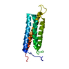
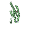
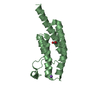
Entry Database : PDB / ID : 6jmuTitle Crystal structure of GIT1/Paxillin complex ARF GTPase-activating protein GIT1 Paxillin Keywords / / / Function / homology Function Domain/homology Component
/ / / / / / / / / / / / / / / / / / / / / / / / / / / / / / / / / / / / / / / / / / / / / / / / / / / / / / / / / / / / / / / / / / / / / / / / / / / / / / / / / / / / / / / / / / / / / / / / / / / / / / / / / / / / / / / / / / / / / / / / / / / / / / / / / / / / / / / / / / / / / / / / Biological species Mus musculus (house mouse)Method / / / Resolution : 2 Å Authors Zhu, J. / Lin, L. / Xia, Y. / Zhang, R. / Zhang, M. Journal : Mol.Cell / Year : 2020Title : GIT/PIX Condensates Are Modular and Ideal for Distinct Compartmentalized Cell Signaling.Authors : Zhu, J. / Zhou, Q. / Xia, Y. / Lin, L. / Li, J. / Peng, M. / Zhang, R. / Zhang, M. History Deposition Mar 13, 2019 Deposition site / Processing site Revision 1.0 May 20, 2020 Provider / Type Revision 1.1 Oct 21, 2020 Group / Category / citation_authorItem _citation.country / _citation.journal_abbrev ... _citation.country / _citation.journal_abbrev / _citation.journal_id_ASTM / _citation.journal_id_CSD / _citation.journal_id_ISSN / _citation.journal_volume / _citation.page_first / _citation.page_last / _citation.pdbx_database_id_DOI / _citation.pdbx_database_id_PubMed / _citation.title / _citation.year Revision 1.2 Nov 22, 2023 Group / Database references / Refinement descriptionCategory chem_comp_atom / chem_comp_bond ... chem_comp_atom / chem_comp_bond / database_2 / pdbx_initial_refinement_model Item / _database_2.pdbx_database_accession
Show all Show less
 Movie
Movie Controller


 Open data
 Basic information
 Components
 Keywords
 Function and homology information
 X-RAY DIFFRACTION /
  Authors
 Citation
 Structure visualization
 Downloads & links
 Other downloads
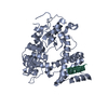


 PDBj
PDBj


 Assembly
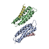


 Mass: 18.015 Da / Num. of mol.: 214 / Source method: isolated from a natural source / Formula: H2O
 Sample preparation
 / Beamline: BL19U1 / Wavelength: 0.97853 Å
 Processing