 Movie
Movie Controller


+ Open data

- Basic information
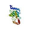
| Entry | Database: PDB / ID: 1x23 | ||||||
|---|---|---|---|---|---|---|---|
| Title | Crystal structure of ubch5c | ||||||
 Components Components | Ubiquitin-conjugating enzyme E2 D3 | ||||||
 Keywords Keywords | LIGASE / Ubiquitin conjugating enzyme / Ubiquitin / E2 | ||||||
| Function / homology |  Function and homology information Function and homology informationSignaling by BMP / (E3-independent) E2 ubiquitin-conjugating enzyme / protein K11-linked ubiquitination / protein K6-linked ubiquitination / : / E2 ubiquitin-conjugating enzyme / ubiquitin conjugating enzyme activity / negative regulation of BMP signaling pathway / protein monoubiquitination / protein autoubiquitination ...Signaling by BMP / (E3-independent) E2 ubiquitin-conjugating enzyme / protein K11-linked ubiquitination / protein K6-linked ubiquitination / : / E2 ubiquitin-conjugating enzyme / ubiquitin conjugating enzyme activity / negative regulation of BMP signaling pathway / protein monoubiquitination / protein autoubiquitination / protein K48-linked ubiquitination / protein modification process / TICAM1, RIP1-mediated IKK complex recruitment / IKK complex recruitment mediated by RIP1 / PINK1-PRKN Mediated Mitophagy / Negative regulators of DDX58/IFIH1 signaling / Peroxisomal protein import / Downregulation of SMAD2/3:SMAD4 transcriptional activity / Regulation of TNFR1 signaling / Inactivation of CSF3 (G-CSF) signaling / Oxygen-dependent proline hydroxylation of Hypoxia-inducible Factor Alpha / protein polyubiquitination / ubiquitin-protein transferase activity / ubiquitin protein ligase activity / Antigen processing: Ubiquitination & Proteasome degradation / E3 ubiquitin ligases ubiquitinate target proteins / Neddylation / ubiquitin-dependent protein catabolic process / proteasome-mediated ubiquitin-dependent protein catabolic process / endosome membrane / protein ubiquitination / DNA repair / apoptotic process / negative regulation of transcription by RNA polymerase II / extracellular exosome / nucleoplasm / ATP binding / nucleus / plasma membrane / cytosol Similarity search - Function | ||||||
| Biological species |  Homo sapiens (human) Homo sapiens (human) | ||||||
| Method |  X-RAY DIFFRACTION / SYNCHROTRON / MOLECULAR REPLACEMENT / Resolution: 1.85 Å X-RAY DIFFRACTION / SYNCHROTRON / MOLECULAR REPLACEMENT / Resolution: 1.85 Å | ||||||
 Authors Authors | Nakanishi, M. / Teshima, N. / Mizushima, T. / Murata, S. / Tanaka, K. / Yamane, T. | ||||||
 Citation Citation | Journal: To be Published Title: Crystal structure of ubch5c Authors: Nakanishi, M. / Teshima, N. / Mizushima, T. / Murata, S. / Tanaka, K. / Yamane, T. | ||||||
| History |
|
- Structure visualization
| Structure viewer | Molecule: MolmilJmol/JSmol |
|---|
- Downloads & links
-Download
| PDBx/mmCIF format | 1x23.cif.gz | 145 KB | Display | PDBx/mmCIF format |
|---|---|---|---|---|
| PDB format | pdb1x23.ent.gz | 114.2 KB | Display | PDB format |
| PDBx/mmJSON format | 1x23.json.gz | Tree view | PDBx/mmJSON format | |
| Others |  Other downloads Other downloads |
-Validation report
| Arichive directory | https://data.pdbj.org/pub/pdb/validation_reports/x2/1x23ftp://data.pdbj.org/pub/pdb/validation_reports/x2/1x23 | HTTPS FTP |
|---|
-Related structure data
| Related structure data | 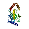 1qcqS S: Starting model for refinement |
|---|---|
| Similar structure data |


-Links
 PDBj
PDBj


- Assembly
| Deposited unit | 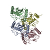
| ||||||||||||||||||||||||||||||
|---|---|---|---|---|---|---|---|---|---|---|---|---|---|---|---|---|---|---|---|---|---|---|---|---|---|---|---|---|---|---|---|
| 1 | 
| ||||||||||||||||||||||||||||||
| 2 | 
| ||||||||||||||||||||||||||||||
| 3 | 
| ||||||||||||||||||||||||||||||
| 4 | 
| ||||||||||||||||||||||||||||||
| Unit cell |
| ||||||||||||||||||||||||||||||
| Noncrystallographic symmetry (NCS) | NCS domain:
NCS domain segments: Component-ID: 1 / Ens-ID: 1 / Beg auth comp-ID: MET / Beg label comp-ID: MET / End auth comp-ID: MET / End label comp-ID: MET / Refine code: 5 / Auth seq-ID: 1 - 147 / Label seq-ID: 9 - 155
|
-Components
| #1: Protein | Mass: 17490.996 Da / Num. of mol.: 4 Source method: isolated from a genetically manipulated source Source: (gene. exp.) Homo sapiens (human) / Plasmid: pGEX6P-1 / Production host:  #2: Water | ChemComp-HOH / |  Mass: 18.015 Da / Num. of mol.: 677 / Source method: isolated from a natural source / Formula: H2O Mass: 18.015 Da / Num. of mol.: 677 / Source method: isolated from a natural source / Formula: H2OHas protein modification | Y | |
|---|
-Experimental details
-Experiment
| Experiment | Method: X-RAY DIFFRACTION / Number of used crystals: 1 |
|---|
- Sample preparation
| Crystal | Density Matthews: 2 Å3/Da / Density % sol: 38.1 % |
|---|---|
| Crystal grow | Temperature: 288 K / Method: vapor diffusion, hanging drop / pH: 7.5 Details: 0.1M HEPES-Na, 10% iso-propanol, 20% PEG4000, pH 7.5, VAPOR DIFFUSION, HANGING DROP, temperature 288K |
-Data collection
| Diffraction | Mean temperature: 100 K |
|---|---|
| Diffraction source | Source: SYNCHROTRON / Site: SPring-8  / Beamline: BL44XU / Wavelength: 0.9 Å / Beamline: BL44XU / Wavelength: 0.9 Å |
| Detector | Type: Bruker DIP-6040 / Detector: CCD / Date: Nov 27, 2004 |
| Radiation | Protocol: SINGLE WAVELENGTH / Monochromatic (M) / Laue (L): M / Scattering type: x-ray |
| Radiation wavelength | Wavelength: 0.9 Å / Relative weight: 1 |
| Reflection | Resolution: 1.8→100 Å / Num. obs: 46077 / % possible obs: 93.9 % / Observed criterion σ(I): 0 / Redundancy: 2.8 % / Biso Wilson estimate: 21.7 Å2 / Rmerge(I) obs: 0.04 / Net I/σ(I): 20.9 |
| Reflection shell | Resolution: 1.8→1.86 Å / Rmerge(I) obs: 0.249 / % possible all: 83.8 |
- Processing
| Software |
| ||||||||||||||||||||||||||||||||||||||||||||||||||||||||||||||||||||||||||||||||||||||||||||||||||||||
|---|---|---|---|---|---|---|---|---|---|---|---|---|---|---|---|---|---|---|---|---|---|---|---|---|---|---|---|---|---|---|---|---|---|---|---|---|---|---|---|---|---|---|---|---|---|---|---|---|---|---|---|---|---|---|---|---|---|---|---|---|---|---|---|---|---|---|---|---|---|---|---|---|---|---|---|---|---|---|---|---|---|---|---|---|---|---|---|---|---|---|---|---|---|---|---|---|---|---|---|---|---|---|---|
| Refinement | Method to determine structure: MOLECULAR REPLACEMENT Starting model: PDB ENTRY 1QCQ Resolution: 1.85→20 Å / Cor.coef. Fo:Fc: 0.951 / Cor.coef. Fo:Fc free: 0.902 / SU B: 4.156 / SU ML: 0.132 / Cross valid method: THROUGHOUT / ESU R: 0.207 / ESU R Free: 0.199 / Stereochemistry target values: MAXIMUM LIKELIHOOD / Details: HYDROGENS HAVE BEEN ADDED IN THE RIDING POSITIONS
| ||||||||||||||||||||||||||||||||||||||||||||||||||||||||||||||||||||||||||||||||||||||||||||||||||||||
| Solvent computation | Ion probe radii: 0.8 Å / Shrinkage radii: 0.8 Å / VDW probe radii: 1.2 Å / Solvent model: MASK | ||||||||||||||||||||||||||||||||||||||||||||||||||||||||||||||||||||||||||||||||||||||||||||||||||||||
| Displacement parameters | Biso mean: 28.5 Å2
| ||||||||||||||||||||||||||||||||||||||||||||||||||||||||||||||||||||||||||||||||||||||||||||||||||||||
| Refinement step | Cycle: LAST / Resolution: 1.85→20 Å
| ||||||||||||||||||||||||||||||||||||||||||||||||||||||||||||||||||||||||||||||||||||||||||||||||||||||
| Refine LS restraints |
| ||||||||||||||||||||||||||||||||||||||||||||||||||||||||||||||||||||||||||||||||||||||||||||||||||||||
| Refine LS restraints NCS | Ens-ID: 1 / Refine-ID: X-RAY DIFFRACTION
| ||||||||||||||||||||||||||||||||||||||||||||||||||||||||||||||||||||||||||||||||||||||||||||||||||||||
| LS refinement shell | Resolution: 1.85→1.898 Å / Total num. of bins used: 20
|
