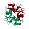
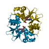
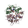
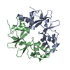
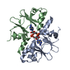
- PDB-2uv7: Crystal Structure of a CBS domain pair from the regulatory gamma1... -
+
Open data
ID or keywords:
Loading...
-
Basic information
Entry
Database: PDB / ID: 2uv7
Title
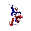
Crystal Structure of a CBS domain pair from the regulatory gamma1 subunit of human AMPK in complex with AMP
Components
5'-AMP-ACTIVATED PROTEIN KINASE SUBUNIT GAMMA-1
Keywords
TRANSFERASE / CBS DOMAIN / LIPID SYNTHESIS / FATTY ACID BIOSYNTHESIS / AMPK GAMMA1 SUBUNIT CBS 3 PLUS 4 AMP REGULATORY SUBUNIT. R TRANSFERASE
Function / homology
Function and homology information
AMP-activated protein kinase activity / Lipophagy / regulation of carbon utilization / cAMP-dependent protein kinase regulator activity / import into nucleus / Energy dependent regulation of mTOR by LKB1-AMPK / nucleotide-activated protein kinase complex / protein kinase regulator activity / Activation of PPARGC1A (PGC-1alpha) by phosphorylation / regulation of glycolytic process ...AMP-activated protein kinase activity / Lipophagy / regulation of carbon utilization / cAMP-dependent protein kinase regulator activity / import into nucleus / Energy dependent regulation of mTOR by LKB1-AMPK / nucleotide-activated protein kinase complex / protein kinase regulator activity / Activation of PPARGC1A (PGC-1alpha) by phosphorylation / regulation of glycolytic process / cAMP-dependent protein kinase activity / AMP binding / Macroautophagy / cellular response to glucose starvation / Activation of AMPK downstream of NMDARs / cellular response to nutrient levels / positive regulation of gluconeogenesis / TP53 Regulates Metabolic Genes / Translocation of SLC2A4 (GLUT4) to the plasma membrane / ADP binding / fatty acid biosynthetic process / spermatogenesis / Regulation of TP53 Activity through Phosphorylation / protein phosphorylation / regulation of cell cycle / protein kinase binding / protein-containing complex binding / signal transduction / nucleoplasm / ATP binding / membrane / nucleus / cytoplasm / cytosol Similarity search - Function
: / CBS-domain / CBS-domain / Domain in cystathionine beta-synthase and other proteins. / CBS domain superfamily / CBS domain / CBS domain / CBS domain profile. / Roll / Alpha Beta Similarity search - Domain/homology
Method to determine structure: OTHER / Resolution: 2→34.94 Å / Cor.coef. Fo:Fc: 0.964 / Cor.coef. Fo:Fc free: 0.938 / SU B: 4.411 / SU ML: 0.124 / Cross valid method: THROUGHOUT / ESU R: 0.197 / ESU R Free: 0.181 / Stereochemistry target values: MAXIMUM LIKELIHOOD / Details: HYDROGENS HAVE BEEN ADDED IN THE RIDING POSITIONS.
Rfactor
Num. reflection
% reflection
Selection details
Rfree
0.23
483
5.1 %
RANDOM
Rwork
0.158
-
-
-
obs
0.162
9026
99.8 %
-
Solvent computation
Ion probe radii: 0.8 Å / Shrinkage radii: 0.8 Å / VDW probe radii: 1.2 Å / Solvent model: MASK
 Movie
Movie Controller

 Yorodumi
 Open data
 Basic information
 Components
 Keywords
 Function and homology information
 HOMO SAPIENS (human)
 X-RAY DIFFRACTION / OTHER / Resolution: 2 Å
  Authors
 Citation
 Structure visualization
 Downloads & links
 Other downloads
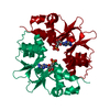


 PDBj
PDBj


 Assembly
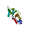
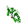


 Mass: 347.221 Da / Num. of mol.: 1 / Source method: obtained synthetically / Formula: C10H14N5O7P / Comment: AMP*YM
 Mass: 18.015 Da / Num. of mol.: 201 / Source method: isolated from a natural source / Formula: H2O
 Sample preparation
 Processing