[English] 日本語
Yorodumi
- PDB-5ks7: Crystal structure of Listeria monocytogenes OpuCA CBS domain dime... -

+
Open data


ID or keywords:

Loading...

-
Basic information

Entry
Database: PDB / ID: 5ks7
TitleCrystal structure of Listeria monocytogenes OpuCA CBS domain dimer in complex with cyclic-di-AMP
ComponentsCarnitine transport ATP-binding protein OpuCA
Keywordshydrolase/hydrolase inhibitor / CBS domain / ABC transporter / Cyclic-di-AMP / Osmoregulation / hydrolase-hydrolase inhibitor complex
Function / homology
Function and homology information


ABC-type quaternary amine transporter / ABC-type quaternary ammonium compound transporting activity / glycine betaine transport / ATP binding / membrane
Similarity search - Function
Glycine betaine transport ATP-binding subunit / CBS-domain / CBS-domain / Domain in cystathionine beta-synthase and other proteins. / CBS domain / CBS domain / CBS domain profile. / ABC transporter-like, conserved site / ABC transporters family signature. / ABC transporter ...Glycine betaine transport ATP-binding subunit / CBS-domain / CBS-domain / Domain in cystathionine beta-synthase and other proteins. / CBS domain / CBS domain / CBS domain profile. / ABC transporter-like, conserved site / ABC transporters family signature. / ABC transporter / ABC transporter-like, ATP-binding domain / ATP-binding cassette, ABC transporter-type domain profile. / ATPases associated with a variety of cellular activities / AAA+ ATPase domain / Roll / P-loop containing nucleoside triphosphate hydrolase / Alpha Beta
Similarity search - Domain/homology
Chem-2BA / Carnitine transport ATP-binding protein OpuCA
Similarity search - Component
Biological speciesListeria monocytogenes (bacteria)
MethodX-RAY DIFFRACTION / MOLECULAR REPLACEMENT / Resolution: 2.9 Å
AuthorsChoi, P.H. / Tong, L.
Funding support United States, 2items
OrganizationGrant numberCountry
National Institutes of Health/National Institute Of Allergy and Infectious Diseases (NIH/NIAID)AI116669 United States
National Institutes of Health/Office of the DirectorOD012018 United States
CitationJournal: Mol.Microbiol. / Year: 2016
Title: Cyclic di-AMP targets the cystathionine beta-synthase domain of the osmolyte transporter OpuC.
Authors: Huynh, T.N. / Choi, P.H. / Sureka, K. / Ledvina, H.E. / Campillo, J. / Tong, L. / Woodward, J.J.
History
DepositionJul 7, 2016Deposition site: RCSB / Processing site: RCSB
Revision 1.0Jul 20, 2016Provider: repository / Type: Initial release
Revision 1.1Oct 19, 2016Group: Database references
Revision 1.2Mar 7, 2018Group: Data collection / Database references / Derived calculations
Category: citation / diffrn_source / pdbx_struct_oper_list
Item: _citation.journal_id_CSD / _diffrn_source.source / _pdbx_struct_oper_list.symmetry_operation
Revision 1.3Mar 23, 2022Group: Author supporting evidence / Database references ...Author supporting evidence / Database references / Derived calculations / Structure summary
Category: chem_comp / database_2 ...chem_comp / database_2 / entity / pdbx_audit_support / pdbx_entity_nonpoly
Item: _chem_comp.name / _database_2.pdbx_DOI ..._chem_comp.name / _database_2.pdbx_DOI / _database_2.pdbx_database_accession / _entity.pdbx_description / _pdbx_entity_nonpoly.name
Revision 1.4Apr 3, 2024Group: Data collection / Refinement description
Category: chem_comp_atom / chem_comp_bond / pdbx_initial_refinement_model

-
Structure visualization

Structure viewerMolecule:
MolmilJmol/JSmol

Downloads & links

-
Assembly

Deposited unit
A: Carnitine transport ATP-binding protein OpuCA
B: Carnitine transport ATP-binding protein OpuCA
hetero molecules


Theoretical massNumber of molelcules
Total (without water)29,2733
Polymers28,6152
Non-polymers6581
Water0
1


  • Idetical with deposited unit
  • defined by author&software
TypeNameSymmetry operationNumber
identity operation1_555x,y,z1
Buried area2930 Å2
ΔGint-14 kcal/mol
Surface area12670 Å2
MethodPISA
Unit cell
Length a, b, c (Å)52.342, 43.861, 68.841
Angle α, β, γ (deg.)90.00, 94.94, 90.00
Int Tables number4
Space group name H-MP1211

-
Components

#1: Protein Carnitine transport ATP-binding protein OpuCA


Mass: 14307.512 Da / Num. of mol.: 2 / Fragment: unp residues 247-372
Source method: isolated from a genetically manipulated source
Source: (gene. exp.) Listeria monocytogenes (bacteria) / Gene: opuCA / Production host: Escherichia coli (E. coli)
References: UniProt: Q9KHT9, quaternary-amine-transporting ATPase
#2: Chemical ChemComp-2BA / (2R,3R,3aS,5R,7aR,9R,10R,10aS,12R,14aR)-2,9-bis(6-amino-9H-purin-9-yl)octahydro-2H,7H-difuro[3,2-d:3',2'-j][1,3,7,9,2,8 ]tetraoxadiphosphacyclododecine-3,5,10,12-tetrol 5,12-dioxide / bis-(3',5')-cyclic-dimeric-Adenosine-monophosphate


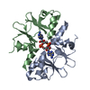
Mass: 658.412 Da / Num. of mol.: 1 / Source method: obtained synthetically / Formula: C20H24N10O12P2

-
Experimental details

-
Experiment

ExperimentMethod: X-RAY DIFFRACTION / Number of used crystals: 1

-
Sample preparation

CrystalDensity Matthews: 2.75 Å3/Da / Density % sol: 55.29 %
Crystal growTemperature: 293 K / Method: vapor diffusion, sitting drop / pH: 9
Details: 0.1M bicine (pH 9.0), 3% (w/v) PEG 20,000, and 2% (v/v) 1,4-dioxane

-
Data collection

DiffractionMean temperature: 100 K
Diffraction sourceSource: ROTATING ANODE / Type: RIGAKU MICROMAX-003 / Wavelength: 1.54 Å
DetectorType: RIGAKU SATURN 944 / Detector: CCD / Date: Sep 10, 2015
RadiationProtocol: SINGLE WAVELENGTH / Monochromatic (M) / Laue (L): M / Scattering type: x-ray
Radiation wavelengthWavelength: 1.54 Å / Relative weight: 1
ReflectionResolution: 2.9→50 Å / Num. obs: 6932 / % possible obs: 97.3 % / Observed criterion σ(I): -3 / Redundancy: 2.5 % / Rmerge(I) obs: 0.115 / Net I/σ(I): 7.9
Reflection shellResolution: 2.9→3 Å / Redundancy: 2 % / Rmerge(I) obs: 0.33 / Mean I/σ(I) obs: 2.2 / % possible all: 89.3

-
Processing

Software
NameVersionClassification
REFMAC5.7.0029refinement
DENZOdata reduction
SCALEPACKdata scaling
PHASERphasing
RefinementMethod to determine structure: MOLECULAR REPLACEMENT
Starting model: AtCBSX1

Resolution: 2.9→43.39 Å / Cor.coef. Fo:Fc: 0.922 / Cor.coef. Fo:Fc free: 0.876 / SU B: 21.025 / SU ML: 0.414 / Cross valid method: THROUGHOUT / ESU R Free: 0.525 / Details: HYDROGENS HAVE BEEN ADDED IN THE RIDING POSITIONS
RfactorNum. reflection% reflectionSelection details
Rfree0.3231 327 4.7 %RANDOM
Rwork0.26572 ---
obs0.26857 6598 97.29 %-
Solvent computationIon probe radii: 0.8 Å / Shrinkage radii: 0.8 Å / VDW probe radii: 1.2 Å
Displacement parametersBiso mean: 26.001 Å2
Baniso -1Baniso -2Baniso -3
1--2.38 Å2-0 Å21.37 Å2
2--1.97 Å2-0 Å2
3---0.29 Å2
Refinement stepCycle: LAST / Resolution: 2.9→43.39 Å
ProteinNucleic acidLigandSolventTotal
Num. atoms1928 0 44 0 1972
Refine LS restraints
Refine-IDTypeDev idealDev ideal targetNumber
X-RAY DIFFRACTIONr_bond_refined_d0.0130.021994
X-RAY DIFFRACTIONr_bond_other_d0.0020.022006
X-RAY DIFFRACTIONr_angle_refined_deg1.6712.012714
X-RAY DIFFRACTIONr_angle_other_deg0.80834596
X-RAY DIFFRACTIONr_dihedral_angle_1_deg8.3695243
X-RAY DIFFRACTIONr_dihedral_angle_2_deg42.05624.81581
X-RAY DIFFRACTIONr_dihedral_angle_3_deg20.64315374
X-RAY DIFFRACTIONr_dihedral_angle_4_deg20.1811514
X-RAY DIFFRACTIONr_chiral_restr0.0790.2346
X-RAY DIFFRACTIONr_gen_planes_refined0.0060.022157
X-RAY DIFFRACTIONr_gen_planes_other0.0010.02403
X-RAY DIFFRACTIONr_nbd_refined
X-RAY DIFFRACTIONr_nbd_other
X-RAY DIFFRACTIONr_nbtor_refined
X-RAY DIFFRACTIONr_nbtor_other
X-RAY DIFFRACTIONr_xyhbond_nbd_refined
X-RAY DIFFRACTIONr_xyhbond_nbd_other
X-RAY DIFFRACTIONr_metal_ion_refined
X-RAY DIFFRACTIONr_metal_ion_other
X-RAY DIFFRACTIONr_symmetry_vdw_refined
X-RAY DIFFRACTIONr_symmetry_vdw_other
X-RAY DIFFRACTIONr_symmetry_hbond_refined
X-RAY DIFFRACTIONr_symmetry_hbond_other
X-RAY DIFFRACTIONr_symmetry_metal_ion_refined
X-RAY DIFFRACTIONr_symmetry_metal_ion_other
X-RAY DIFFRACTIONr_mcbond_it
X-RAY DIFFRACTIONr_mcbond_other
X-RAY DIFFRACTIONr_mcangle_it
X-RAY DIFFRACTIONr_mcangle_other
X-RAY DIFFRACTIONr_scbond_it
X-RAY DIFFRACTIONr_scbond_other
X-RAY DIFFRACTIONr_scangle_it
X-RAY DIFFRACTIONr_scangle_other
X-RAY DIFFRACTIONr_long_range_B_refined
X-RAY DIFFRACTIONr_long_range_B_other
X-RAY DIFFRACTIONr_rigid_bond_restr
X-RAY DIFFRACTIONr_sphericity_free
X-RAY DIFFRACTIONr_sphericity_bonded
LS refinement shellResolution: 2.896→3.053 Å / Total num. of bins used: 10
RfactorNum. reflection% reflection
Rfree0.371 40 -
Rwork0.325 866 -
obs--89.88 %

+
About Yorodumi

-
News

-
Feb 9, 2022. New format data for meta-information of EMDB entries

New format data for meta-information of EMDB entries

  • Version 3 of the EMDB header file is now the official format.
  • The previous official version 1.9 will be removed from the archive.

Related info.:EMDB header

External links:wwPDB to switch to version 3 of the EMDB data model

-
Aug 12, 2020. Covid-19 info

Covid-19 info

URL: https://pdbj.org/emnavi/covid19.php

New page: Covid-19 featured information page in EM Navigator.

Related info.:Covid-19 info / Mar 5, 2020. Novel coronavirus structure data

+
Mar 5, 2020. Novel coronavirus structure data

Novel coronavirus structure data

Related info.:Yorodumi Speices / Aug 12, 2020. Covid-19 info

External links:COVID-19 featured content - PDBj / Molecule of the Month (242):Coronavirus Proteases

+
Jan 31, 2019. EMDB accession codes are about to change! (news from PDBe EMDB page)

EMDB accession codes are about to change! (news from PDBe EMDB page)

  • The allocation of 4 digits for EMDB accession codes will soon come to an end. Whilst these codes will remain in use, new EMDB accession codes will include an additional digit and will expand incrementally as the available range of codes is exhausted. The current 4-digit format prefixed with “EMD-” (i.e. EMD-XXXX) will advance to a 5-digit format (i.e. EMD-XXXXX), and so on. It is currently estimated that the 4-digit codes will be depleted around Spring 2019, at which point the 5-digit format will come into force.
  • The EM Navigator/Yorodumi systems omit the EMD- prefix.

Related info.:Q: What is EMD? / ID/Accession-code notation in Yorodumi/EM Navigator

External links:EMDB Accession Codes are Changing Soon! / Contact to PDBj

+
Jul 12, 2017. Major update of PDB

Major update of PDB

  • wwPDB released updated PDB data conforming to the new PDBx/mmCIF dictionary.
  • This is a major update changing the version number from 4 to 5, and with Remediation, in which all the entries are updated.
  • In this update, many items about electron microscopy experimental information are reorganized (e.g. em_software).
  • Now, EM Navigator and Yorodumi are based on the updated data.

External links:wwPDB Remediation / Enriched Model Files Conforming to OneDep Data Standards Now Available in the PDB FTP Archive

-
Yorodumi

Thousand views of thousand structures

  • Yorodumi is a browser for structure data from EMDB, PDB, SASBDB, etc.
  • This page is also the successor to EM Navigator detail page, and also detail information page/front-end page for Omokage search.
  • The word "yorodu" (or yorozu) is an old Japanese word meaning "ten thousand". "mi" (miru) is to see.

Related info.:EMDB / PDB / SASBDB / Comparison of 3 databanks / Yorodumi Search / Aug 31, 2016. New EM Navigator & Yorodumi / Yorodumi Papers / Jmol/JSmol / Function and homology information / Changes in new EM Navigator and Yorodumi

Read more