 Movie
Movie Controller

[English] 日本語
 Yorodumi
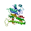
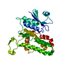
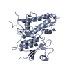
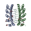
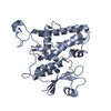
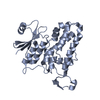
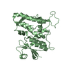
- PDB-6zn8: Crystal structure of the H. influenzae VapXD toxin-antitoxin complex -
+ Open data

- Basic information
| Entry | Database: PDB / ID: 6zn8 | |||||||||
|---|---|---|---|---|---|---|---|---|---|---|
| Title | Crystal structure of the H. influenzae VapXD toxin-antitoxin complex | |||||||||
 Components Components |
| |||||||||
 Keywords Keywords | TOXIN / Toxin-antitoxin / VapXD / RNase / Nucleic-Acid Binding Protein / Hydrolase | |||||||||
| Function / homology | Protein of unknown function DUF5397 / Family of unknown function (DUF5397) / Endoribonuclease VapD / nuclease activity / Hydrolases; Acting on ester bonds / hydrolase activity / RNA binding / VapX / Endoribonuclease VapD Function and homology information Function and homology information | |||||||||
| Biological species |  Haemophilus influenzae (bacteria) Haemophilus influenzae (bacteria) | |||||||||
| Method |  X-RAY DIFFRACTION / SYNCHROTRON / SAD / Resolution: 3.211 Å X-RAY DIFFRACTION / SYNCHROTRON / SAD / Resolution: 3.211 Å | |||||||||
 Authors Authors | Bertelsen, M.B. / Senissar, M. / Nielsen, M.H. / Bisiak, F. / Cunha, M.V. / Molinaro, A.L. / Daines, D.A. / Brodersen, D.E. | |||||||||
| Funding support |  Denmark, 2items Denmark, 2items
| |||||||||
 Citation Citation | Journal: Structure / Year: 2021 Title: Structural Basis for Toxin Inhibition in the VapXD Toxin-Antitoxin System. Authors: Bertelsen, M.B. / Senissar, M. / Nielsen, M.H. / Bisiak, F. / Cunha, M.V. / Molinaro, A.L. / Daines, D.A. / Brodersen, D.E. | |||||||||
| History |
|
- Structure visualization
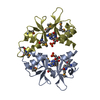
| Structure viewer | Molecule: MolmilJmol/JSmol |
|---|
- Downloads & links
-Download
| PDBx/mmCIF format | 6zn8.cif.gz | 110.6 KB | Display | PDBx/mmCIF format |
|---|---|---|---|---|
| PDB format | pdb6zn8.ent.gz | 86.8 KB | Display | PDB format |
| PDBx/mmJSON format | 6zn8.json.gz | Tree view | PDBx/mmJSON format | |
| Others |  Other downloads Other downloads |
-Validation report
| Arichive directory | https://data.pdbj.org/pub/pdb/validation_reports/zn/6zn8ftp://data.pdbj.org/pub/pdb/validation_reports/zn/6zn8 | HTTPS FTP |
|---|
-Related structure data


-Links
 PDBj
PDBj- Assembly
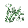
| Deposited unit | 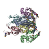
| ||||||||
|---|---|---|---|---|---|---|---|---|---|
| 1 | 
| ||||||||
| 2 | 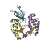
| ||||||||
| Unit cell |
|
-Components
| #1: Protein | Mass: 11915.762 Da / Num. of mol.: 4 Source method: isolated from a genetically manipulated source Source: (gene. exp.) Haemophilus influenzae (strain 86-028NP) (bacteria)Gene: vapD, NTHI0577 / Cell line (production host): B834 (DE3) / Production host: References: UniProt: Q4QN95, Hydrolases; Acting on ester bonds #2: Protein | Mass: 9089.575 Da / Num. of mol.: 2 Source method: isolated from a genetically manipulated source Source: (gene. exp.) Haemophilus influenzae (strain 86-028NP) (bacteria)Gene: vapX, NTHI0578 / Cell line (production host): B834 (DE3) / Production host: Has ligand of interest | N | Has protein modification | Y | |
|---|
-Experimental details
-Experiment
| Experiment | Method: X-RAY DIFFRACTION / Number of used crystals: 1 |
|---|
- Sample preparation
| Crystal | Density Matthews: 5.36 Å3/Da / Density % sol: 77.03 % |
|---|---|
| Crystal grow | Temperature: 292 K / Method: vapor diffusion, sitting drop / pH: 6.5 / Details: 1.6 M ammonium sulfate, 10% (v/v) 1,4-dioxane |
-Data collection
| Diffraction | Mean temperature: 100 K / Serial crystal experiment: N | ||||||||||||||||||||||||
|---|---|---|---|---|---|---|---|---|---|---|---|---|---|---|---|---|---|---|---|---|---|---|---|---|---|
| Diffraction source | Source: SYNCHROTRON / Site: ESRF  / Beamline: ID29 / Wavelength: 0.97903 Å / Beamline: ID29 / Wavelength: 0.97903 Å | ||||||||||||||||||||||||
| Detector | Type: DECTRIS PILATUS 6M-F / Detector: PIXEL / Date: Apr 6, 2018 | ||||||||||||||||||||||||
| Radiation | Protocol: SINGLE WAVELENGTH / Monochromatic (M) / Laue (L): M / Scattering type: x-ray | ||||||||||||||||||||||||
| Radiation wavelength | Wavelength: 0.97903 Å / Relative weight: 1 | ||||||||||||||||||||||||
| Reflection | Resolution: 3.21→46.88 Å / Num. obs: 23799 / % possible obs: 99.8 % / Redundancy: 7.3 % / Biso Wilson estimate: 100.67 Å2 / CC1/2: 0.994 / Rmerge(I) obs: 0.171 / Rpim(I) all: 0.067 / Rrim(I) all: 0.185 / Net I/σ(I): 8.2 | ||||||||||||||||||||||||
| Reflection shell | Diffraction-ID: 1
|
- Processing
| Software |
| ||||||||||||||||||||||||||||||||||||||||||||||||||||||||||||||||||||||||||||||||||||||||||||||||||||||||||||
|---|---|---|---|---|---|---|---|---|---|---|---|---|---|---|---|---|---|---|---|---|---|---|---|---|---|---|---|---|---|---|---|---|---|---|---|---|---|---|---|---|---|---|---|---|---|---|---|---|---|---|---|---|---|---|---|---|---|---|---|---|---|---|---|---|---|---|---|---|---|---|---|---|---|---|---|---|---|---|---|---|---|---|---|---|---|---|---|---|---|---|---|---|---|---|---|---|---|---|---|---|---|---|---|---|---|---|---|---|---|
| Refinement | Method to determine structure: SAD / Resolution: 3.211→46.876 Å / SU ML: 0.48 / Cross valid method: THROUGHOUT / σ(F): 1.36 / Phase error: 29.34
| ||||||||||||||||||||||||||||||||||||||||||||||||||||||||||||||||||||||||||||||||||||||||||||||||||||||||||||
| Solvent computation | Shrinkage radii: 0.8 Å / VDW probe radii: 1.1 Å | ||||||||||||||||||||||||||||||||||||||||||||||||||||||||||||||||||||||||||||||||||||||||||||||||||||||||||||
| Displacement parameters | Biso max: 154.25 Å2 / Biso mean: 85.5947 Å2 / Biso min: 46.7 Å2 | ||||||||||||||||||||||||||||||||||||||||||||||||||||||||||||||||||||||||||||||||||||||||||||||||||||||||||||
| Refinement step | Cycle: final / Resolution: 3.211→46.876 Å
| ||||||||||||||||||||||||||||||||||||||||||||||||||||||||||||||||||||||||||||||||||||||||||||||||||||||||||||
| Refine LS restraints |
| ||||||||||||||||||||||||||||||||||||||||||||||||||||||||||||||||||||||||||||||||||||||||||||||||||||||||||||
| LS refinement shell | Refine-ID: X-RAY DIFFRACTION / Rfactor Rfree error: 0
|
