 Movie
Movie Controller

[English] 日本語
 Yorodumi
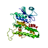
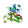
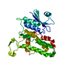
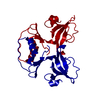
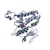
- PDB-1k0m: Crystal structure of a soluble monomeric form of CLIC1 at 1.4 ang... -
+ Open data

- Basic information
| Entry | Database: PDB / ID: 1k0m | ||||||
|---|---|---|---|---|---|---|---|
| Title | Crystal structure of a soluble monomeric form of CLIC1 at 1.4 angstroms | ||||||
 Components Components | CHLORIDE INTRACELLULAR CHANNEL PROTEIN 1 | ||||||
 Keywords Keywords | METAL TRANSPORT / glutathione-S-tranferase superfamily / Chloride Ion Channel | ||||||
| Function / homology |  Function and homology information Function and homology informationglutathione dehydrogenase (ascorbate) / glutathione dehydrogenase (ascorbate) activity / Oxidoreductases; Acting on a sulfur group of donors / chloride transport / chloride channel activity / brush border / positive regulation of osteoblast differentiation / chloride channel complex / regulation of mitochondrial membrane potential / platelet aggregation ...glutathione dehydrogenase (ascorbate) / glutathione dehydrogenase (ascorbate) activity / Oxidoreductases; Acting on a sulfur group of donors / chloride transport / chloride channel activity / brush border / positive regulation of osteoblast differentiation / chloride channel complex / regulation of mitochondrial membrane potential / platelet aggregation / nuclear envelope / blood microparticle / nuclear membrane / vesicle / cadherin binding / perinuclear region of cytoplasm / signal transduction / endoplasmic reticulum / mitochondrion / : / extracellular exosome / membrane / nucleus / plasma membrane / cytoplasm Similarity search - Function | ||||||
| Biological species |  Homo sapiens (human) Homo sapiens (human) | ||||||
| Method |  X-RAY DIFFRACTION / SYNCHROTRON / MIR / Resolution: 1.4 Å X-RAY DIFFRACTION / SYNCHROTRON / MIR / Resolution: 1.4 Å | ||||||
 Authors Authors | Harrop, S.J. / DeMaere, M.Z. / Fairlie, W.D. / Reztsova, T. / Valenzuela, S.M. / Mazzanti, M. / Tonini, R. / Qiu, M.R. / Jankova, L. / Warton, K. ...Harrop, S.J. / DeMaere, M.Z. / Fairlie, W.D. / Reztsova, T. / Valenzuela, S.M. / Mazzanti, M. / Tonini, R. / Qiu, M.R. / Jankova, L. / Warton, K. / Bauskin, A.R. / Wu, W.M. / Pankhurst, S. / Campbell, T.J. / Breit, S.N. / Curmi, P.M.G. | ||||||
 Citation Citation | Journal: J.Biol.Chem. / Year: 2001 Title: Crystal structure of a soluble form of the intracellular chloride ion channel CLIC1 (NCC27) at 1.4-A resolution. Authors: Harrop, S.J. / DeMaere, M.Z. / Fairlie, W.D. / Reztsova, T. / Valenzuela, S.M. / Mazzanti, M. / Tonini, R. / Qiu, M.R. / Jankova, L. / Warton, K. / Bauskin, A.R. / Wu, W.M. / Pankhurst, S. / ...Authors: Harrop, S.J. / DeMaere, M.Z. / Fairlie, W.D. / Reztsova, T. / Valenzuela, S.M. / Mazzanti, M. / Tonini, R. / Qiu, M.R. / Jankova, L. / Warton, K. / Bauskin, A.R. / Wu, W.M. / Pankhurst, S. / Campbell, T.J. / Breit, S.N. / Curmi, P.M. | ||||||
| History |
|
- Structure visualization
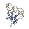
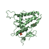
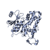
| Structure viewer | Molecule: MolmilJmol/JSmol |
|---|
- Downloads & links
-Download
| PDBx/mmCIF format | 1k0m.cif.gz | 214.9 KB | Display | PDBx/mmCIF format |
|---|---|---|---|---|
| PDB format | pdb1k0m.ent.gz | 173.3 KB | Display | PDB format |
| PDBx/mmJSON format | 1k0m.json.gz | Tree view | PDBx/mmJSON format | |
| Others |  Other downloads Other downloads |
-Validation report
| Arichive directory | https://data.pdbj.org/pub/pdb/validation_reports/k0/1k0mftp://data.pdbj.org/pub/pdb/validation_reports/k0/1k0m | HTTPS FTP |
|---|
-Related structure data


-Links
 PDBj
PDBj

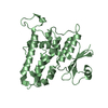
- Assembly
| Deposited unit | 
| ||||||||
|---|---|---|---|---|---|---|---|---|---|
| 1 | 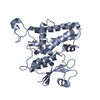
| ||||||||
| 2 | 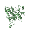
| ||||||||
| Unit cell |
| ||||||||
| Details | SOLUBLE MONOMERIC FORM |
-Components
| #1: Protein | Mass: 26881.605 Da / Num. of mol.: 2 / Fragment: CLIC1 / Mutation: E151G Source method: isolated from a genetically manipulated source Source: (gene. exp.) Homo sapiens (human) / Plasmid: pGEX-4T-1 / Production host:  #2: Water | ChemComp-HOH / |  Mass: 18.015 Da / Num. of mol.: 656 / Source method: isolated from a natural source / Formula: H2O Mass: 18.015 Da / Num. of mol.: 656 / Source method: isolated from a natural source / Formula: H2O |
|---|
-Experimental details
-Experiment
| Experiment | Method: X-RAY DIFFRACTION / Number of used crystals: 1 |
|---|
- Sample preparation
| Crystal | Density Matthews: 2.08 Å3/Da / Density % sol: 40.74 % | ||||||||||||||||||||||||||||||||||||
|---|---|---|---|---|---|---|---|---|---|---|---|---|---|---|---|---|---|---|---|---|---|---|---|---|---|---|---|---|---|---|---|---|---|---|---|---|---|
| Crystal grow | Temperature: 277 K / Method: vapor diffusion, sitting drop / pH: 5 Details: PEG MME 5K, AMMONIUM SULPHATE, SODIUM ACETATE, pH 5.0, VAPOR DIFFUSION, SITTING DROP, temperature 277K | ||||||||||||||||||||||||||||||||||||
| Crystal grow | *PLUS Temperature: 4 ℃ / Method: vapor diffusion | ||||||||||||||||||||||||||||||||||||
| Components of the solutions | *PLUS
|
-Data collection
| Diffraction | Mean temperature: 100 K |
|---|---|
| Diffraction source | Source: SYNCHROTRON / Site: SSRL  / Beamline: BL9-1 / Wavelength: 0.98 Å / Beamline: BL9-1 / Wavelength: 0.98 Å |
| Detector | Type: MARRESEARCH / Detector: IMAGE PLATE / Date: Jul 15, 1998 / Details: MONOCHROMATOR |
| Radiation | Monochromator: YES / Protocol: SINGLE WAVELENGTH / Monochromatic (M) / Laue (L): M / Scattering type: x-ray |
| Radiation wavelength | Wavelength: 0.98 Å / Relative weight: 1 |
| Reflection | Resolution: 1.4→87 Å / Num. all: 81905 / Num. obs: 81905 / % possible obs: 94.7 % / Observed criterion σ(F): 0 / Observed criterion σ(I): 0 / Redundancy: 5.2 % / Biso Wilson estimate: 14 Å2 / Rmerge(I) obs: 0.022 / Rsym value: 0.022 / Net I/σ(I): 28.9 |
| Reflection shell | Resolution: 1.4→1.42 Å / Redundancy: 4.8 % / Rmerge(I) obs: 0.098 / Mean I/σ(I) obs: 9.3 / Num. unique all: 4039 / Rsym value: 0.098 / % possible all: 94.2 |
| Reflection | *PLUS Lowest resolution: 87 Å / Num. obs: 81729 / % possible obs: 94.1 % / Num. measured all: 259290 / Rmerge(I) obs: 0.03 |
| Reflection shell | *PLUS Highest resolution: 1.4 Å / % possible obs: 94 % / Rmerge(I) obs: 0.08 / Mean I/σ(I) obs: 6.5 |
- Processing
| Software |
| |||||||||||||||||||||||||
|---|---|---|---|---|---|---|---|---|---|---|---|---|---|---|---|---|---|---|---|---|---|---|---|---|---|---|
| Refinement | Method to determine structure: MIR / Resolution: 1.4→87.71 Å / Isotropic thermal model: anisotropic / Cross valid method: THROUGHOUT / σ(F): 0 / σ(I): 0 / Stereochemistry target values: Engh & Huber Details: TLS parameters refined for the N and C domains in each monomer
| |||||||||||||||||||||||||
| Displacement parameters | Biso mean: 10.755 Å2
| |||||||||||||||||||||||||
| Refinement step | Cycle: LAST / Resolution: 1.4→87.71 Å
| |||||||||||||||||||||||||
| Refine LS restraints |
| |||||||||||||||||||||||||
| LS refinement shell | Highest resolution: 1.4 Å / Rfactor Rfree: 0.177 / Rfactor Rwork: 0.127 / Total num. of bins used: 20 | |||||||||||||||||||||||||
| Software | *PLUS Name: REFMAC / Version: 5 / Classification: refinement | |||||||||||||||||||||||||
| Refinement | *PLUS σ(F): 0 / % reflection Rfree: 5 % / Rfactor obs: 0.138 | |||||||||||||||||||||||||
| Solvent computation | *PLUS | |||||||||||||||||||||||||
| Displacement parameters | *PLUS | |||||||||||||||||||||||||
| Refine LS restraints | *PLUS
|
