- PDB-2qyz: Crystal structure of the uncharacterized protein CTC02137 from Cl... -
+
Open data
ID or keywords:
Loading...
-
Basic information
Entry
Database: PDB / ID: 2qyz
Title
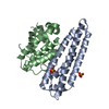
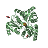
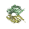
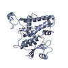
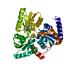
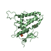
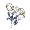
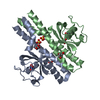
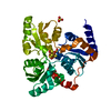
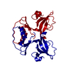
Crystal structure of the uncharacterized protein CTC02137 from Clostridium tetani E88
Components
Uncharacterized protein
Keywords
STRUCTURAL GENOMICS / UNKNOWN FUNCTION / Clostridium tetani E88 / PSI-2 / Protein Structure Initiative / New York SGX Research Center for Structural Genomics / NYSGXRC
Function / homology
Function and homology information
ctc02137 like domains / YopX-like domains / Conserved hypothetical protein CHP1671 / YopX-like, C-terminal / YopX protein / YopX protein / Dna Ligase; domain 1 / SH3 type barrels. / Roll / 2-Layer Sandwich ...ctc02137 like domains / YopX-like domains / Conserved hypothetical protein CHP1671 / YopX-like, C-terminal / YopX protein / YopX protein / Dna Ligase; domain 1 / SH3 type barrels. / Roll / 2-Layer Sandwich / Mainly Beta / Alpha Beta Similarity search - Domain/homology
BIOMOLECULE: 1 SEE REMARK 350 FOR THE AUTHOR PROVIDED AND PROGRAM GENERATED ASSEMBLY INFORMATION ... BIOMOLECULE: 1 SEE REMARK 350 FOR THE AUTHOR PROVIDED AND PROGRAM GENERATED ASSEMBLY INFORMATION FOR THE STRUCTURE IN THIS ENTRY. THE REMARK MAY ALSO PROVIDE INFORMATION ON BURIED SURFACE AREA. THE DIMER IS DISULFIDE-LINKED THROUGH CYS-92.
Protocol: SINGLE WAVELENGTH / Monochromatic (M) / Laue (L): M / Scattering type: x-ray
Radiation wavelength
Wavelength: 0.979 Å / Relative weight: 1
Reflection
Redundancy: 4.9 % / Av σ(I) over netI: 18.3 / Number: 81550 / Rmerge(I) obs: 0.035 / Χ2: 1.14 / D res high: 2.04 Å / D res low: 50 Å / Num. obs: 16582 / % possible obs: 89.3
Diffraction reflection shell
Highest resolution (Å)
Lowest resolution (Å)
% possible obs (%)
ID
Rmerge(I) obs
Chi squared
Redundancy
4.39
50
96.3
1
0.024
1.784
5.3
3.49
4.39
96.6
1
0.036
2.088
5.3
3.05
3.49
100
1
0.036
1.244
5.7
2.77
3.05
100
1
0.048
0.992
5.7
2.57
2.77
99.9
1
0.063
0.809
5.7
2.42
2.57
100
1
0.09
0.681
5.5
2.3
2.42
98.9
1
0.109
0.618
4.7
2.2
2.3
89.3
1
0.196
1.369
3.5
2.11
2.2
71.6
1
0.172
0.523
3
2.04
2.11
39.6
1
0.183
0.509
2.6
Reflection
Resolution: 2.04→50 Å / Num. obs: 16582 / % possible obs: 89.3 % / Redundancy: 4.9 % / Rmerge(I) obs: 0.035 / Χ2: 1.143 / Net I/σ(I): 18.3
Reflection shell
Resolution (Å)
Redundancy (%)
Rmerge(I) obs
Num. unique all
Χ2
Diffraction-ID
% possible all
2.04-2.11
2.6
0.183
722
0.509
1
39.6
2.11-2.2
3
0.172
1326
0.523
1
71.6
2.2-2.3
3.5
0.196
1656
1.369
1
89.3
2.3-2.42
4.7
0.109
1847
0.618
1
98.9
2.42-2.57
5.5
0.09
1866
0.681
1
100
2.57-2.77
5.7
0.063
1834
0.809
1
99.9
2.77-3.05
5.7
0.048
1848
0.992
1
100
3.05-3.49
5.7
0.036
1872
1.244
1
100
3.49-4.39
5.3
0.036
1796
2.088
1
96.6
4.39-50
5.3
0.024
1815
1.784
1
96.3
-
Processing
Software
Name
Version
Classification
NB
DENZO
datareduction
SCALEPACK
datascaling
REFMAC
refinement
PDB_EXTRACT
3
dataextraction
CBASS
datacollection
SHELXS
phasing
Refinement
Resolution: 2.04→19.7 Å / Cor.coef. Fo:Fc: 0.968 / Cor.coef. Fo:Fc free: 0.938 / SU B: 22.616 / SU ML: 0.242 / TLS residual ADP flag: LIKELY RESIDUAL / Cross valid method: THROUGHOUT / σ(F): 0 / ESU R: 0.212 / ESU R Free: 0.199 / Stereochemistry target values: MAXIMUM LIKELIHOOD Details: The Bijvoet differences were used for phasing. HYDROGENS HAVE BEEN ADDED IN THE RIDING POSITIONS
Rfactor
Num. reflection
% reflection
Selection details
Rfree
0.259
435
4.8 %
RANDOM
Rwork
0.191
-
-
-
obs
0.194
9007
92.09 %
-
Solvent computation
Ion probe radii: 0.8 Å / Shrinkage radii: 0.8 Å / VDW probe radii: 1.2 Å / Solvent model: BABINET MODEL WITH MASK
Displacement parameters
Biso mean: 54.382 Å2
Baniso -1
Baniso -2
Baniso -3
1-
0.21 Å2
0.1 Å2
0 Å2
2-
-
0.21 Å2
0 Å2
3-
-
-
-0.31 Å2
Refinement step
Cycle: LAST / Resolution: 2.04→19.7 Å
Protein
Nucleic acid
Ligand
Solvent
Total
Num. atoms
1044
0
0
67
1111
Refine LS restraints
Refine-ID
Type
Dev ideal
Dev ideal target
Number
X-RAY DIFFRACTION
r_bond_refined_d
0.021
0.022
1066
X-RAY DIFFRACTION
r_angle_refined_deg
2.008
1.956
1438
X-RAY DIFFRACTION
r_dihedral_angle_1_deg
8.538
5
128
X-RAY DIFFRACTION
r_dihedral_angle_2_deg
42.195
25.614
57
X-RAY DIFFRACTION
r_dihedral_angle_3_deg
19.268
15
202
X-RAY DIFFRACTION
r_dihedral_angle_4_deg
25.263
15
5
X-RAY DIFFRACTION
r_chiral_restr
0.132
0.2
155
X-RAY DIFFRACTION
r_gen_planes_refined
0.008
0.02
809
X-RAY DIFFRACTION
r_nbd_refined
0.243
0.2
412
X-RAY DIFFRACTION
r_nbtor_refined
0.322
0.2
718
X-RAY DIFFRACTION
r_xyhbond_nbd_refined
0.157
0.2
57
X-RAY DIFFRACTION
r_symmetry_vdw_refined
0.222
0.2
62
X-RAY DIFFRACTION
r_symmetry_hbond_refined
0.165
0.2
9
X-RAY DIFFRACTION
r_mcbond_it
1.807
1.5
657
X-RAY DIFFRACTION
r_mcangle_it
5.987
20
1031
X-RAY DIFFRACTION
r_scbond_it
11.645
20
476
X-RAY DIFFRACTION
r_scangle_it
5.213
4.5
406
LS refinement shell
Resolution: 2.04→2.095 Å / Total num. of bins used: 20
Rfactor
Num. reflection
% reflection
Rfree
0.532
21
-
Rwork
0.326
291
-
all
-
312
-
obs
-
-
45.55 %
Refinement TLS params.
Method: refined / Origin x: 0.9503 Å / Origin y: 5.2605 Å / Origin z: 36.9146 Å
11
12
13
21
22
23
31
32
33
T
-0.0044 Å2
-0.1084 Å2
0.0072 Å2
-
-0.1403 Å2
-0.0111 Å2
-
-
-0.0676 Å2
L
1.3446 °2
-0.138 °2
-0.4823 °2
-
1.9553 °2
-1.3127 °2
-
-
4.7512 °2
S
0.0854 Å °
-0.2281 Å °
0.0447 Å °
0.1964 Å °
-0.2359 Å °
0.0842 Å °
0.0135 Å °
0.1044 Å °
0.1505 Å °
+
About Yorodumi
-
News
-
Feb 9, 2022. New format data for meta-information of EMDB entries
New format data for meta-information of EMDB entries
Version 3 of the EMDB header file is now the official format.
The previous official version 1.9 will be removed from the archive.
In the structure databanks used in Yorodumi, some data are registered as the other names, "COVID-19 virus" and "2019-nCoV". Here are the details of the virus and the list of structure data.
Jan 31, 2019. EMDB accession codes are about to change! (news from PDBe EMDB page)
EMDB accession codes are about to change! (news from PDBe EMDB page)
The allocation of 4 digits for EMDB accession codes will soon come to an end. Whilst these codes will remain in use, new EMDB accession codes will include an additional digit and will expand incrementally as the available range of codes is exhausted. The current 4-digit format prefixed with “EMD-” (i.e. EMD-XXXX) will advance to a 5-digit format (i.e. EMD-XXXXX), and so on. It is currently estimated that the 4-digit codes will be depleted around Spring 2019, at which point the 5-digit format will come into force.
The EM Navigator/Yorodumi systems omit the EMD- prefix.
Related info.:Q: What is EMD? / ID/Accession-code notation in Yorodumi/EM Navigator
Yorodumi is a browser for structure data from EMDB, PDB, SASBDB, etc.
This page is also the successor to EM Navigator detail page, and also detail information page/front-end page for Omokage search.
The word "yorodu" (or yorozu) is an old Japanese word meaning "ten thousand". "mi" (miru) is to see.
Related info.:EMDB / PDB / SASBDB / Comparison of 3 databanks / Yorodumi Search / Aug 31, 2016. New EM Navigator & Yorodumi / Yorodumi Papers / Jmol/JSmol / Function and homology information / Changes in new EM Navigator and Yorodumi
 Movie
Movie Controller

 Yorodumi
 Open data
 Basic information
 Components
 Keywords
 Function and homology information
 Clostridium tetani E88 (bacteria)
 X-RAY DIFFRACTION /
  Authors
 Citation
 Structure visualization
 Downloads & links
 Other downloads
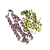
 PDBj
PDBj Assembly
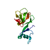
 Mass: 18.015 Da / Num. of mol.: 67 / Source method: isolated from a natural source / Formula: H2O
 Sample preparation
 / Beamline: X29A / Wavelength: 0.979 Å
 Processing