 Movie
Movie Controller

[English] 日本語
 Yorodumi
- PDB-3ik4: CRYSTAL STRUCTURE OF mandelate racemase/muconate lactonizing prot... -
+ Open data

- Basic information
| Entry | Database: PDB / ID: 3ik4 | ||||||
|---|---|---|---|---|---|---|---|
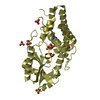
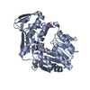
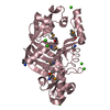
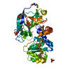
| Title | CRYSTAL STRUCTURE OF mandelate racemase/muconate lactonizing protein from Herpetosiphon aurantiacus | ||||||
 Components Components | Mandelate racemase/muconate lactonizing protein | ||||||
 Keywords Keywords | ISOMERASE / STRUCTURAL GENOMICS / ENOLASE / EPIMERASE / PSI-2 / PROTEIN STRUCTURE INITIATIVE / NEW YORK SGX RESEARCH CENTER FOR STRUCTURAL GENOMICS / NYSGXRC | ||||||
| Function / homology |  Function and homology information Function and homology informationracemase and epimerase activity, acting on amino acids and derivatives / Isomerases; Racemases and epimerases; Acting on amino acids and derivatives / racemase and epimerase activity / peptide metabolic process / magnesium ion binding Similarity search - Function | ||||||
| Biological species |  Herpetosiphon aurantiacus ATCC 23779 (bacteria) Herpetosiphon aurantiacus ATCC 23779 (bacteria) | ||||||
| Method |  X-RAY DIFFRACTION / SYNCHROTRON / molecular replacement / Resolution: 2.1 Å X-RAY DIFFRACTION / SYNCHROTRON / molecular replacement / Resolution: 2.1 Å | ||||||
 Authors Authors | Patskovsky, Y. / Toro, R. / Dickey, M. / Iizuka, M. / Sauder, J.M. / Gerlt, J.A. / Burley, S.K. / Almo, S.C. / New York SGX Research Center for Structural Genomics (NYSGXRC) | ||||||
 Citation Citation | Journal: Proc.Natl.Acad.Sci.USA / Year: 2012 Title: Homology models guide discovery of diverse enzyme specificities among dipeptide epimerases in the enolase superfamily. Authors: Lukk, T. / Sakai, A. / Kalyanaraman, C. / Brown, S.D. / Imker, H.J. / Song, L. / Fedorov, A.A. / Fedorov, E.V. / Toro, R. / Hillerich, B. / Seidel, R. / Patskovsky, Y. / Vetting, M.W. / ...Authors: Lukk, T. / Sakai, A. / Kalyanaraman, C. / Brown, S.D. / Imker, H.J. / Song, L. / Fedorov, A.A. / Fedorov, E.V. / Toro, R. / Hillerich, B. / Seidel, R. / Patskovsky, Y. / Vetting, M.W. / Nair, S.K. / Babbitt, P.C. / Almo, S.C. / Gerlt, J.A. / Jacobson, M.P. | ||||||
| History |
|
- Structure visualization
| Structure viewer | Molecule: MolmilJmol/JSmol |
|---|
- Downloads & links
-Download
| PDBx/mmCIF format | 3ik4.cif.gz | 267.4 KB | Display | PDBx/mmCIF format |
|---|---|---|---|---|
| PDB format | pdb3ik4.ent.gz | 215.9 KB | Display | PDB format |
| PDBx/mmJSON format | 3ik4.json.gz | Tree view | PDBx/mmJSON format | |
| Others |  Other downloads Other downloads |
-Validation report
| Arichive directory | https://data.pdbj.org/pub/pdb/validation_reports/ik/3ik4ftp://data.pdbj.org/pub/pdb/validation_reports/ik/3ik4 | HTTPS FTP |
|---|
-Related structure data
| Related structure data | 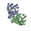 3ijiC  3ijlC 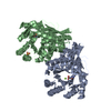 3ijqC  3jvaC  3jw7C  3jzuC  3k1gC  3kumC  3q45C  3q4dC  3r0kC  3r0uC  3r10C  3r11C 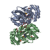 3r1zC  3ritC  3ro6C  1jpmS S: Starting model for refinement C: citing same article ( |
|---|---|
| Similar structure data | |
| Other databases |


-Links
 PDBj
PDBj
- Assembly
| Deposited unit | 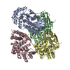
| ||||||||
|---|---|---|---|---|---|---|---|---|---|
| 1 | 
| ||||||||
| 2 | 
| ||||||||
| 3 | 
| ||||||||
| 4 | 
| ||||||||
| 5 | 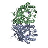
| ||||||||
| 6 | 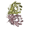
| ||||||||
| Unit cell |
|
-Components
| #1: Protein | Mass: 37963.426 Da / Num. of mol.: 4 Source method: isolated from a genetically manipulated source Source: (gene. exp.) Herpetosiphon aurantiacus ATCC 23779 (bacteria)Gene: Haur_1114 / Production host: #2: Chemical | ChemComp-K /   Mass: 39.098 Da / Num. of mol.: 4 / Source method: obtained synthetically / Formula: K Mass: 39.098 Da / Num. of mol.: 4 / Source method: obtained synthetically / Formula: K#3: Chemical |   Mass: 92.094 Da / Num. of mol.: 2 / Source method: obtained synthetically / Formula: C3H8O3 Mass: 92.094 Da / Num. of mol.: 2 / Source method: obtained synthetically / Formula: C3H8O3#4: Water | ChemComp-HOH / |  Mass: 18.015 Da / Num. of mol.: 237 / Source method: isolated from a natural source / Formula: H2O Mass: 18.015 Da / Num. of mol.: 237 / Source method: isolated from a natural source / Formula: H2O |
|---|
-Experimental details
-Experiment
| Experiment | Method: X-RAY DIFFRACTION / Number of used crystals: 1 |
|---|
- Sample preparation
| Crystal | Density Matthews: 2.06 Å3/Da / Density % sol: 40.26 % |
|---|---|
| Crystal grow | Method: vapor diffusion, sitting drop / pH: 7.5 Details: 150MM POTASSIUM BROMIDE, PH 7.5, 30% PEG2000 MME, 10% GLYCEROL, VAPOR DIFFUSION, SITTING DROP, TEMPERATURE 294K |
-Data collection
| Diffraction | Mean temperature: 90 K |
|---|---|
| Diffraction source | Source: SYNCHROTRON / Site: NSLS  / Beamline: X29A / Wavelength: 1.081 / Beamline: X29A / Wavelength: 1.081 |
| Detector | Type: ADSC QUANTUM 315 / Detector: CCD / Date: Jul 23, 2009 / Details: MIRRORS |
| Radiation | Protocol: SINGLE WAVELENGTH / Monochromatic (M) / Laue (L): M / Scattering type: x-ray |
| Radiation wavelength | Wavelength: 1.081 Å / Relative weight: 1 |
| Reflection | Resolution: 2.07→50 Å / Num. obs: 75228 / % possible obs: 98.9 % / Observed criterion σ(I): -5 / Redundancy: 4.1 % / Biso Wilson estimate: 43.053 Å2 / Rsym value: 0.063 / Net I/σ(I): 7.9 |
| Reflection shell | Resolution: 2.07→2.14 Å / Redundancy: 3.7 % / Rmerge(I) obs: 0.78 / Mean I/σ(I) obs: 0.9 / % possible all: 91.2 |
- Processing
| Software |
| ||||||||||||||||||||||||||||||||||||||||||||||||||||||||||||||||||||||||||||||||||||||||||||||||||||||||||||||||||||||||||||||||||||||||||||||||||||||||||||||||||||||||||
|---|---|---|---|---|---|---|---|---|---|---|---|---|---|---|---|---|---|---|---|---|---|---|---|---|---|---|---|---|---|---|---|---|---|---|---|---|---|---|---|---|---|---|---|---|---|---|---|---|---|---|---|---|---|---|---|---|---|---|---|---|---|---|---|---|---|---|---|---|---|---|---|---|---|---|---|---|---|---|---|---|---|---|---|---|---|---|---|---|---|---|---|---|---|---|---|---|---|---|---|---|---|---|---|---|---|---|---|---|---|---|---|---|---|---|---|---|---|---|---|---|---|---|---|---|---|---|---|---|---|---|---|---|---|---|---|---|---|---|---|---|---|---|---|---|---|---|---|---|---|---|---|---|---|---|---|---|---|---|---|---|---|---|---|---|---|---|---|---|---|---|---|
| Refinement | Method to determine structure: molecular replacement Starting model: 1JPM Resolution: 2.1→20 Å / Cor.coef. Fo:Fc: 0.961 / Cor.coef. Fo:Fc free: 0.935 / SU B: 20.061 / SU ML: 0.247 / TLS residual ADP flag: LIKELY RESIDUAL / Cross valid method: THROUGHOUT / ESU R: 0.29 / ESU R Free: 0.229 / Stereochemistry target values: MAXIMUM LIKELIHOOD / Details: HYDROGENS HAVE BEEN ADDED IN THE RIDING POSITIONS
| ||||||||||||||||||||||||||||||||||||||||||||||||||||||||||||||||||||||||||||||||||||||||||||||||||||||||||||||||||||||||||||||||||||||||||||||||||||||||||||||||||||||||||
| Solvent computation | Ion probe radii: 0.8 Å / Shrinkage radii: 0.8 Å / VDW probe radii: 1.4 Å / Solvent model: MASK | ||||||||||||||||||||||||||||||||||||||||||||||||||||||||||||||||||||||||||||||||||||||||||||||||||||||||||||||||||||||||||||||||||||||||||||||||||||||||||||||||||||||||||
| Displacement parameters | Biso mean: 41.917 Å2
| ||||||||||||||||||||||||||||||||||||||||||||||||||||||||||||||||||||||||||||||||||||||||||||||||||||||||||||||||||||||||||||||||||||||||||||||||||||||||||||||||||||||||||
| Refinement step | Cycle: LAST / Resolution: 2.1→20 Å
| ||||||||||||||||||||||||||||||||||||||||||||||||||||||||||||||||||||||||||||||||||||||||||||||||||||||||||||||||||||||||||||||||||||||||||||||||||||||||||||||||||||||||||
| Refine LS restraints |
| ||||||||||||||||||||||||||||||||||||||||||||||||||||||||||||||||||||||||||||||||||||||||||||||||||||||||||||||||||||||||||||||||||||||||||||||||||||||||||||||||||||||||||
| LS refinement shell | Resolution: 2.1→2.154 Å / Total num. of bins used: 20
| ||||||||||||||||||||||||||||||||||||||||||||||||||||||||||||||||||||||||||||||||||||||||||||||||||||||||||||||||||||||||||||||||||||||||||||||||||||||||||||||||||||||||||
| Refinement TLS params. | Method: refined / Refine-ID: X-RAY DIFFRACTION
| ||||||||||||||||||||||||||||||||||||||||||||||||||||||||||||||||||||||||||||||||||||||||||||||||||||||||||||||||||||||||||||||||||||||||||||||||||||||||||||||||||||||||||
| Refinement TLS group |
|
