 Movie
Movie Controller

[English] 日本語
 Yorodumi
- PDB-3hvq: Crystal structure of a complex between Protein Phosphatase 1 alph... -
+ Open data

- Basic information
| Entry | Database: PDB / ID: 3hvq | ||||||
|---|---|---|---|---|---|---|---|
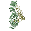
| Title | Crystal structure of a complex between Protein Phosphatase 1 alpha (PP1) and the PP1 binding and PDZ domains of Neurabin | ||||||
 Components Components |
| ||||||
 Keywords Keywords | HYDROLASE/HYDROLASE REGULATOR / PP1 / NEURABIN / SERINE/THREONINE PHOSPHATASE / POST SYNAPTIC DENSITY / GLUTAMETERGIC RECEPTORS / CARBOHYDRATE METABOLISM / CELL CYCLE / CELL DIVISION / GLYCOGEN METABOLISM / HYDROLASE / IRON / MANGANESE / METAL-BINDING / PHOSPHOPROTEIN / PROTEIN PHOSPHATASE / ACTIN-BINDING / CELL JUNCTION / CELL PROJECTION / CYTOSKELETON / DEVELOPMENTAL PROTEIN / DIFFERENTIATION / NEUROGENESIS / NUCLEUS / SYNAPSE / Synaptosome / HYDROLASE-HYDROLASE REGULATOR COMPLEX | ||||||
| Function / homology |  Function and homology information Function and homology informationnegative regulation of spontaneous neurotransmitter secretion / regulation of synapse structural plasticity / regulation of glycogen catabolic process / positive regulation of termination of RNA polymerase II transcription, poly(A)-coupled / postsynaptic actin cytoskeleton organization / positive regulation of long-term synaptic depression / PTW/PP1 phosphatase complex / protein phosphatase type 1 complex / glycogen granule / RNA polymerase II promoter clearance ...negative regulation of spontaneous neurotransmitter secretion / regulation of synapse structural plasticity / regulation of glycogen catabolic process / positive regulation of termination of RNA polymerase II transcription, poly(A)-coupled / postsynaptic actin cytoskeleton organization / positive regulation of long-term synaptic depression / PTW/PP1 phosphatase complex / protein phosphatase type 1 complex / glycogen granule / RNA polymerase II promoter clearance / RNA polymerase II CTD heptapeptide repeat S5 phosphatase activity / cadherin binding involved in cell-cell adhesion / protein phosphatase 1 binding / regulation of filopodium assembly / regulation of translational initiation in response to stress / regulation of actin filament polymerization / regulation of dendritic spine morphogenesis / negative regulation of stress fiber assembly / positive regulation of extrinsic apoptotic signaling pathway in absence of ligand / postsynaptic actin cytoskeleton / cellular response to toxic substance / growth cone lamellipodium / positive regulation of protein kinase activity / regulation of canonical Wnt signaling pathway / protein dephosphorylation / Phosphorylation and nuclear translocation of the CRY:PER:kinase complex / dendritic spine neck / glycogen metabolic process / cortical actin cytoskeleton / branching morphogenesis of an epithelial tube / Triglyceride catabolism / protein-serine/threonine phosphatase / entrainment of circadian clock by photoperiod / regulation of synapse assembly / positive regulation of dendritic spine development / protein serine/threonine phosphatase activity / telomere maintenance in response to DNA damage / phosphatase activity / Maturation of hRSV A proteins / negative regulation of long-term synaptic potentiation / negative regulation of transcription elongation by RNA polymerase II / transition metal ion binding / positive regulation of glycogen biosynthetic process / DARPP-32 events / ribonucleoprotein complex binding / neuron development / phosphoprotein phosphatase activity / lung development / Downregulation of TGF-beta receptor signaling / actin filament organization / filopodium / circadian regulation of gene expression / adherens junction / neuromuscular junction / centriole / positive regulation of transcription elongation by RNA polymerase II / calcium-mediated signaling / sperm end piece / excitatory postsynaptic potential / positive regulation of neuron projection development / regulation of circadian rhythm / response to lead ion / modulation of chemical synaptic transmission / neuron projection development / actin filament binding / lamellipodium / actin cytoskeleton / growth cone / presynapse / GTPase binding / sperm midpiece / dendritic spine / cytoskeleton / transmembrane transporter binding / perikaryon / postsynaptic density / protein stabilization / iron ion binding / protein domain specific binding / cell division / neuronal cell body / dendrite / protein kinase binding / protein-containing complex binding / nucleolus / glutamatergic synapse / endoplasmic reticulum / extracellular exosome / nucleoplasm / identical protein binding / nucleus / plasma membrane / cytoplasm / cytosol Similarity search - Function | ||||||
| Biological species |  Homo sapiens (human) Homo sapiens (human) | ||||||
| Method |  X-RAY DIFFRACTION / SYNCHROTRON / MOLECULAR REPLACEMENT / Resolution: 2.2 Å X-RAY DIFFRACTION / SYNCHROTRON / MOLECULAR REPLACEMENT / Resolution: 2.2 Å | ||||||
 Authors Authors | Critton, D.A. / Ragusa, M.J. / Page, R. / Peti, W. | ||||||
 Citation Citation | Journal: Nat.Struct.Mol.Biol. / Year: 2010 Title: Spinophilin directs protein phosphatase 1 specificity by blocking substrate binding sites. Authors: Ragusa, M.J. / Dancheck, B. / Critton, D.A. / Nairn, A.C. / Page, R. / Peti, W. | ||||||
| History |
|
- Structure visualization
| Structure viewer | Molecule: MolmilJmol/JSmol |
|---|
- Downloads & links
-Download
| PDBx/mmCIF format | 3hvq.cif.gz | 191.5 KB | Display | PDBx/mmCIF format |
|---|---|---|---|---|
| PDB format | pdb3hvq.ent.gz | 148.8 KB | Display | PDB format |
| PDBx/mmJSON format | 3hvq.json.gz | Tree view | PDBx/mmJSON format | |
| Others |  Other downloads Other downloads |
-Validation report
| Arichive directory | https://data.pdbj.org/pub/pdb/validation_reports/hv/3hvqftp://data.pdbj.org/pub/pdb/validation_reports/hv/3hvq | HTTPS FTP |
|---|
-Related structure data
| Related structure data |  3eggSC  3eghC C: citing same article ( S: Starting model for refinement |
|---|---|
| Similar structure data |


-Links
 PDBj
PDBj


- Assembly
| Deposited unit | 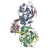
| ||||||||||||||||||||||||||||||||||||||||||||||||||||||||||||||||||||
|---|---|---|---|---|---|---|---|---|---|---|---|---|---|---|---|---|---|---|---|---|---|---|---|---|---|---|---|---|---|---|---|---|---|---|---|---|---|---|---|---|---|---|---|---|---|---|---|---|---|---|---|---|---|---|---|---|---|---|---|---|---|---|---|---|---|---|---|---|---|
| 1 |
| ||||||||||||||||||||||||||||||||||||||||||||||||||||||||||||||||||||
| 2 | 
| ||||||||||||||||||||||||||||||||||||||||||||||||||||||||||||||||||||
| 3 | 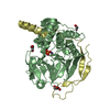
| ||||||||||||||||||||||||||||||||||||||||||||||||||||||||||||||||||||
| Unit cell |
| ||||||||||||||||||||||||||||||||||||||||||||||||||||||||||||||||||||
| Components on special symmetry positions |
| ||||||||||||||||||||||||||||||||||||||||||||||||||||||||||||||||||||
| Noncrystallographic symmetry (NCS) | NCS domain:
NCS domain segments: Component-ID: 1 / Refine code: 5
NCS ensembles :
|
-Components
-Protein , 2 types, 4 molecules ABCD
| #1: Protein | Mass: 37349.844 Da / Num. of mol.: 2 / Fragment: CATALYTIC SUBUNIT Source method: isolated from a genetically manipulated source Source: (gene. exp.) Homo sapiens (human) / Gene: PPP1A, PPP1CA / Plasmid: RP1B / Production host:  References: UniProt: P62136, protein-serine/threonine phosphatase #2: Protein | Mass: 18849.867 Da / Num. of mol.: 2 / Fragment: PP1 BINDING AND PDZ DOMAINS Source method: isolated from a genetically manipulated source Source: (gene. exp.) |
|---|
-Non-polymers , 4 types, 569 molecules 


| #3: Chemical | ChemComp-MN /  Mass: 54.938 Da / Num. of mol.: 4 / Source method: obtained synthetically / Formula: Mn Mass: 54.938 Da / Num. of mol.: 4 / Source method: obtained synthetically / Formula: Mn#4: Chemical |  Mass: 94.971 Da / Num. of mol.: 2 / Source method: obtained synthetically / Formula: PO4 Mass: 94.971 Da / Num. of mol.: 2 / Source method: obtained synthetically / Formula: PO4#5: Chemical |  Mass: 92.094 Da / Num. of mol.: 3 / Source method: obtained synthetically / Formula: C3H8O3 Mass: 92.094 Da / Num. of mol.: 3 / Source method: obtained synthetically / Formula: C3H8O3#6: Water | ChemComp-HOH / | Mass: 18.015 Da / Num. of mol.: 560 / Source method: isolated from a natural source / Formula: H2O |
|---|
-Experimental details
-Experiment
| Experiment | Method: X-RAY DIFFRACTION / Number of used crystals: 1 |
|---|
- Sample preparation
| Crystal | Density Matthews: 2.44 Å3/Da / Density % sol: 49.63 % |
|---|---|
| Crystal grow | Temperature: 277 K / Method: vapor diffusion, sitting drop / pH: 7.9 Details: 0.2 M (NH4)2HPO4, 20% PEG 3350, pH 7.9, VAPOR DIFFUSION, SITTING DROP, temperature 277K |
-Data collection
| Diffraction | Mean temperature: 93 K |
|---|---|
| Diffraction source | Source: SYNCHROTRON / Site: NSLS  / Beamline: X6A / Wavelength: 1 Å / Beamline: X6A / Wavelength: 1 Å |
| Detector | Type: ADSC QUANTUM 270 / Detector: CCD / Date: Dec 5, 2008 Details: DOUBLE CRYSTAL CHANNEL CUT, SI(111), 1M LONG RH COATED TOROIDAL MIRROR FOR VERTICAL AND HORIZONTAL FOCUSING |
| Radiation | Monochromator: SI(111) CHANNEL CUT MONOCHROMATOR / Protocol: SINGLE WAVELENGTH / Monochromatic (M) / Laue (L): M / Scattering type: x-ray |
| Radiation wavelength | Wavelength: 1 Å / Relative weight: 1 |
| Reflection | Resolution: 2.2→20 Å / Num. all: 54834 / Num. obs: 53709 / % possible obs: 97.9 % / Observed criterion σ(F): 0 / Observed criterion σ(I): 0 / Redundancy: 3.3 % / Biso Wilson estimate: 25.5 Å2 / Rsym value: 0.103 / Net I/σ(I): 10.4 |
| Reflection shell | Resolution: 2.2→2.24 Å / Redundancy: 3.4 % / Mean I/σ(I) obs: 5 / Num. unique all: 2731 / Rsym value: 0.263 / % possible all: 97.5 |
- Processing
| Software |
| ||||||||||||||||||||||||||||||||||||||||||||||||||||||||||||||||||||||||||||||||||||||||||||||||||||||||||||||||||||||||||||||||||||||||||||||||||||||||||||||||||||||||||||||||||||||||||||||||||||||||
|---|---|---|---|---|---|---|---|---|---|---|---|---|---|---|---|---|---|---|---|---|---|---|---|---|---|---|---|---|---|---|---|---|---|---|---|---|---|---|---|---|---|---|---|---|---|---|---|---|---|---|---|---|---|---|---|---|---|---|---|---|---|---|---|---|---|---|---|---|---|---|---|---|---|---|---|---|---|---|---|---|---|---|---|---|---|---|---|---|---|---|---|---|---|---|---|---|---|---|---|---|---|---|---|---|---|---|---|---|---|---|---|---|---|---|---|---|---|---|---|---|---|---|---|---|---|---|---|---|---|---|---|---|---|---|---|---|---|---|---|---|---|---|---|---|---|---|---|---|---|---|---|---|---|---|---|---|---|---|---|---|---|---|---|---|---|---|---|---|---|---|---|---|---|---|---|---|---|---|---|---|---|---|---|---|---|---|---|---|---|---|---|---|---|---|---|---|---|---|---|---|---|
| Refinement | Method to determine structure: MOLECULAR REPLACEMENT Starting model: PDB ENTRY 3EGG Resolution: 2.2→20 Å / Cor.coef. Fo:Fc: 0.959 / Cor.coef. Fo:Fc free: 0.926 / SU B: 8.339 / SU ML: 0.113 / TLS residual ADP flag: LIKELY RESIDUAL / Cross valid method: THROUGHOUT / σ(F): 0 / ESU R: 0.202 / ESU R Free: 0.186 / Stereochemistry target values: MAXIMUM LIKELIHOOD / Details: HYDROGENS HAVE BEEN ADDED IN THE RIDING POSITIONS
| ||||||||||||||||||||||||||||||||||||||||||||||||||||||||||||||||||||||||||||||||||||||||||||||||||||||||||||||||||||||||||||||||||||||||||||||||||||||||||||||||||||||||||||||||||||||||||||||||||||||||
| Solvent computation | Ion probe radii: 0.8 Å / Shrinkage radii: 0.8 Å / VDW probe radii: 1.2 Å / Solvent model: MASK | ||||||||||||||||||||||||||||||||||||||||||||||||||||||||||||||||||||||||||||||||||||||||||||||||||||||||||||||||||||||||||||||||||||||||||||||||||||||||||||||||||||||||||||||||||||||||||||||||||||||||
| Displacement parameters | Biso mean: 23.592 Å2
| ||||||||||||||||||||||||||||||||||||||||||||||||||||||||||||||||||||||||||||||||||||||||||||||||||||||||||||||||||||||||||||||||||||||||||||||||||||||||||||||||||||||||||||||||||||||||||||||||||||||||
| Refinement step | Cycle: LAST / Resolution: 2.2→20 Å
| ||||||||||||||||||||||||||||||||||||||||||||||||||||||||||||||||||||||||||||||||||||||||||||||||||||||||||||||||||||||||||||||||||||||||||||||||||||||||||||||||||||||||||||||||||||||||||||||||||||||||
| Refine LS restraints |
| ||||||||||||||||||||||||||||||||||||||||||||||||||||||||||||||||||||||||||||||||||||||||||||||||||||||||||||||||||||||||||||||||||||||||||||||||||||||||||||||||||||||||||||||||||||||||||||||||||||||||
| Refine LS restraints NCS | Dom-ID: 1 / Refine-ID: X-RAY DIFFRACTION
| ||||||||||||||||||||||||||||||||||||||||||||||||||||||||||||||||||||||||||||||||||||||||||||||||||||||||||||||||||||||||||||||||||||||||||||||||||||||||||||||||||||||||||||||||||||||||||||||||||||||||
| LS refinement shell | Resolution: 2.201→2.258 Å / Total num. of bins used: 20
| ||||||||||||||||||||||||||||||||||||||||||||||||||||||||||||||||||||||||||||||||||||||||||||||||||||||||||||||||||||||||||||||||||||||||||||||||||||||||||||||||||||||||||||||||||||||||||||||||||||||||
| Refinement TLS params. | Method: refined / Refine-ID: X-RAY DIFFRACTION
| ||||||||||||||||||||||||||||||||||||||||||||||||||||||||||||||||||||||||||||||||||||||||||||||||||||||||||||||||||||||||||||||||||||||||||||||||||||||||||||||||||||||||||||||||||||||||||||||||||||||||
| Refinement TLS group |
|
