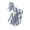
- PDB-3aqp: Crystal structure of SecDF, a translocon-associated membrane prot... -
+
Open data
ID or keywords:
Loading...
-
Basic information
Entry
Database: PDB / ID: 3aqp
Title
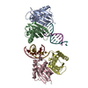
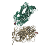
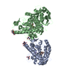
Crystal structure of SecDF, a translocon-associated membrane protein, from Thermus thrmophilus
Components
Probable SecDF protein-export membrane protein
Keywords
MEMBRANE PROTEIN / SECDF / SEC / TRANSLOCON / CELL MEMBRANE / MEMBRANE / PROTEIN TRANSPORT / TRANSLOCATION / TRANSMEMBRANE / TRANSPORT
Function / homology
Function and homology information
protein transport by the Sec complex / intracellular protein transmembrane transport / protein-transporting ATPase activity / protein targeting / plasma membrane Similarity search - Function
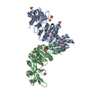
Gyrase A; domain 2 - #200 / Alpha-Beta Plaits - #3220 / Protein-export membrane protein SecF, bacterial / Protein translocase subunit SecD / Protein-export membrane protein SecD/SecF, bacterial / Protein-export membrane protein SecD/SecF/SecDF, conserved site / Protein-export membrane protein SecD/SecF, archaeal and bacterial / : / : / : ...Gyrase A; domain 2 - #200 / Alpha-Beta Plaits - #3220 / Protein-export membrane protein SecF, bacterial / Protein translocase subunit SecD / Protein-export membrane protein SecD/SecF, bacterial / Protein-export membrane protein SecD/SecF/SecDF, conserved site / Protein-export membrane protein SecD/SecF, archaeal and bacterial / : / : / : / : / Protein export membrane protein SecD/SecF, C-terminal / SecD/SecF GG Motif / Protein translocase subunit SecDF, P1 domain, N-terminal / SecDF, P1 head subdomain / Sterol-sensing domain / Gyrase A; domain 2 / Alpha-Beta Plaits / 2-Layer Sandwich / Alpha Beta Similarity search - Domain/homology
In the structure databanks used in Yorodumi, some data are registered as the other names, "COVID-19 virus" and "2019-nCoV". Here are the details of the virus and the list of structure data.
Jan 31, 2019. EMDB accession codes are about to change! (news from PDBe EMDB page)
EMDB accession codes are about to change! (news from PDBe EMDB page)
The allocation of 4 digits for EMDB accession codes will soon come to an end. Whilst these codes will remain in use, new EMDB accession codes will include an additional digit and will expand incrementally as the available range of codes is exhausted. The current 4-digit format prefixed with “EMD-” (i.e. EMD-XXXX) will advance to a 5-digit format (i.e. EMD-XXXXX), and so on. It is currently estimated that the 4-digit codes will be depleted around Spring 2019, at which point the 5-digit format will come into force.
The EM Navigator/Yorodumi systems omit the EMD- prefix.
Related info.:Q: What is EMD? / ID/Accession-code notation in Yorodumi/EM Navigator
Yorodumi is a browser for structure data from EMDB, PDB, SASBDB, etc.
This page is also the successor to EM Navigator detail page, and also detail information page/front-end page for Omokage search.
The word "yorodu" (or yorozu) is an old Japanese word meaning "ten thousand". "mi" (miru) is to see.
Related info.:EMDB / PDB / SASBDB / Comparison of 3 databanks / Yorodumi Search / Aug 31, 2016. New EM Navigator & Yorodumi / Yorodumi Papers / Jmol/JSmol / Function and homology information / Changes in new EM Navigator and Yorodumi
 Movie
Movie Controller

 Yorodumi
 Open data
 Basic information
 Components
 Keywords
 Function and homology information
 Thermus thermophilus (bacteria)
 X-RAY DIFFRACTION /
  Authors
 Citation
 Structure visualization
 Downloads & links
 Other downloads
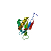


 PDBj
PDBj
 Assembly

 Sample preparation
 / Beamline: BL41XU
 Processing