 Movie
Movie Controller

[English] 日本語
 Yorodumi
- PDB-2bap: Crystal structure of the N-terminal mDia1 Armadillo Repeat Region... -
+ Open data

- Basic information
| Entry | Database: PDB / ID: 2bap | ||||||
|---|---|---|---|---|---|---|---|
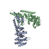
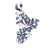
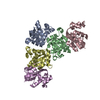
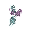
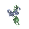
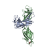
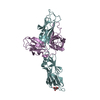
| Title | Crystal structure of the N-terminal mDia1 Armadillo Repeat Region and Dimerisation Domain in complex with the mDia1 autoregulatory domain (DAD) | ||||||
 Components Components | (Diaphanous protein homolog 1) x 2 | ||||||
 Keywords Keywords | SIGNALING PROTEIN / Armadillo Repeats / all helical | ||||||
| Function / homology |  Function and homology information Function and homology informationnegative regulation of neuron projection regeneration / multicellular organismal locomotion / ERBB2 Regulates Cell Motility / RHOF GTPase cycle / RHOC GTPase cycle / : / neuron projection retraction / actin nucleation / RHOB GTPase cycle / RHOA GTPase cycle ...negative regulation of neuron projection regeneration / multicellular organismal locomotion / ERBB2 Regulates Cell Motility / RHOF GTPase cycle / RHOC GTPase cycle / : / neuron projection retraction / actin nucleation / RHOB GTPase cycle / RHOA GTPase cycle / protein localization to microtubule / RHO GTPases Activate Formins / profilin binding / cellular response to histamine / regulation of release of sequestered calcium ion into cytosol / regulation of microtubule-based process / axon midline choice point recognition / regulation of cytoskeleton organization / brush border / ephrin receptor signaling pathway / synaptic vesicle endocytosis / actin filament polymerization / Neutrophil degranulation / cytoskeleton organization / actin filament / sensory perception of sound / brain development / small GTPase binding / ruffle membrane / neuron projection development / mitotic spindle / intracellular protein localization / regulation of cell shape / presynapse / actin cytoskeleton organization / actin binding / gene expression / transmembrane transporter binding / neuron projection / positive regulation of cell migration / centrosome / identical protein binding / nucleus / cytoplasm Similarity search - Function | ||||||
| Biological species |  | ||||||
| Method |  X-RAY DIFFRACTION / SYNCHROTRON / MOLECULAR REPLACEMENT / Resolution: 3.3 Å X-RAY DIFFRACTION / SYNCHROTRON / MOLECULAR REPLACEMENT / Resolution: 3.3 Å | ||||||
 Authors Authors | Lammers, M. / Rose, R. / Scrima, A. / Wittinghofer, A. | ||||||
 Citation Citation | Journal: Embo J. / Year: 2005 Title: The regulation of mDia1 by autoinhibition and its release by Rho*GTP. Authors: Lammers, M. / Rose, R. / Scrima, A. / Wittinghofer, A. | ||||||
| History |
|
- Structure visualization
| Structure viewer | Molecule: MolmilJmol/JSmol |
|---|
- Downloads & links
-Download
| PDBx/mmCIF format | 2bap.cif.gz | 132.7 KB | Display | PDBx/mmCIF format |
|---|---|---|---|---|
| PDB format | pdb2bap.ent.gz | 103.9 KB | Display | PDB format |
| PDBx/mmJSON format | 2bap.json.gz | Tree view | PDBx/mmJSON format | |
| Others |  Other downloads Other downloads |
-Validation report
| Arichive directory | https://data.pdbj.org/pub/pdb/validation_reports/ba/2bapftp://data.pdbj.org/pub/pdb/validation_reports/ba/2bap | HTTPS FTP |
|---|
-Related structure data
| Related structure data | 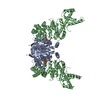 1z2cS S: Starting model for refinement |
|---|---|
| Similar structure data |

-Links
 PDBj
PDBj


- Assembly
| Deposited unit | 
| ||||||||||||||||||||||||||||||||||||||||||||||||||||||||||||||||||||
|---|---|---|---|---|---|---|---|---|---|---|---|---|---|---|---|---|---|---|---|---|---|---|---|---|---|---|---|---|---|---|---|---|---|---|---|---|---|---|---|---|---|---|---|---|---|---|---|---|---|---|---|---|---|---|---|---|---|---|---|---|---|---|---|---|---|---|---|---|---|
| 1 | 
| ||||||||||||||||||||||||||||||||||||||||||||||||||||||||||||||||||||
| 2 | 
| ||||||||||||||||||||||||||||||||||||||||||||||||||||||||||||||||||||
| Unit cell |
| ||||||||||||||||||||||||||||||||||||||||||||||||||||||||||||||||||||
| Noncrystallographic symmetry (NCS) | NCS domain:
NCS domain segments: Component-ID: 1 / Refine code: 4
NCS ensembles :
|
-Components
| #1: Protein | Mass: 36534.020 Da / Num. of mol.: 2 / Fragment: mDia1 N-terminal regulatory domain Source method: isolated from a genetically manipulated source Source: (gene. exp.)  #2: Protein | Mass: 6460.428 Da / Num. of mol.: 2 / Fragment: mDia1 autoregulatory domain, DAD Source method: isolated from a genetically manipulated source Source: (gene. exp.) |
|---|
-Experimental details
-Experiment
| Experiment | Method: X-RAY DIFFRACTION / Number of used crystals: 1 |
|---|
- Sample preparation
| Crystal | Density Matthews: 3.39 Å3/Da / Density % sol: 63.74 % |
|---|---|
| Crystal grow | Temperature: 293 K / Method: vapor diffusion, hanging drop / pH: 7.1 Details: 3.7 M NaFormiate pH 7.1, 100 mM HEPES pH 7.1, 4% (w/v) PEG5000-MME, VAPOR DIFFUSION, HANGING DROP, temperature 293K |
-Data collection
| Diffraction | Mean temperature: 100 K |
|---|---|
| Diffraction source | Source: SYNCHROTRON / Site: SLS  / Beamline: X10SA / Wavelength: 0.95 Å / Beamline: X10SA / Wavelength: 0.95 Å |
| Detector | Type: MARRESEARCH / Detector: CCD / Date: May 28, 2005 |
| Radiation | Monochromator: LN2 cooled fixed-exit Si(111) monochromator / Protocol: SINGLE WAVELENGTH / Monochromatic (M) / Laue (L): M / Scattering type: x-ray |
| Radiation wavelength | Wavelength: 0.95 Å / Relative weight: 1 |
| Reflection | Resolution: 3.3→20 Å / Num. obs: 18490 / % possible obs: 81.9 % / Rmerge(I) obs: 0.141 / Net I/σ(I): 16.79 |
| Reflection shell | Resolution: 3.3→3.4 Å / % possible obs: 0 % / Num. measured obs: 0 / Num. unique obs: 0 / % possible all: 100 |
- Processing
| Software |
| ||||||||||||||||||||||||||||||||||||||||||||||||||||||||||||||||||||||||||||||||||||||||||
|---|---|---|---|---|---|---|---|---|---|---|---|---|---|---|---|---|---|---|---|---|---|---|---|---|---|---|---|---|---|---|---|---|---|---|---|---|---|---|---|---|---|---|---|---|---|---|---|---|---|---|---|---|---|---|---|---|---|---|---|---|---|---|---|---|---|---|---|---|---|---|---|---|---|---|---|---|---|---|---|---|---|---|---|---|---|---|---|---|---|---|---|
| Refinement | Method to determine structure: MOLECULAR REPLACEMENT Starting model: pdb entry 1Z2C Resolution: 3.3→19.98 Å / Cor.coef. Fo:Fc: 0.901 / Cor.coef. Fo:Fc free: 0.839 / SU B: 38.004 / SU ML: 0.621 / Cross valid method: THROUGHOUT / σ(F): 0 / ESU R Free: 0.65 / Stereochemistry target values: MAXIMUM LIKELIHOOD / Details: HYDROGENS HAVE BEEN ADDED IN THE RIDING POSITIONS
| ||||||||||||||||||||||||||||||||||||||||||||||||||||||||||||||||||||||||||||||||||||||||||
| Solvent computation | Ion probe radii: 0.8 Å / Shrinkage radii: 0.8 Å / VDW probe radii: 1.2 Å / Solvent model: MASK | ||||||||||||||||||||||||||||||||||||||||||||||||||||||||||||||||||||||||||||||||||||||||||
| Displacement parameters | Biso mean: 92.189 Å2 | ||||||||||||||||||||||||||||||||||||||||||||||||||||||||||||||||||||||||||||||||||||||||||
| Refinement step | Cycle: LAST / Resolution: 3.3→19.98 Å
| ||||||||||||||||||||||||||||||||||||||||||||||||||||||||||||||||||||||||||||||||||||||||||
| Refine LS restraints |
| ||||||||||||||||||||||||||||||||||||||||||||||||||||||||||||||||||||||||||||||||||||||||||
| Refine LS restraints NCS | Dom-ID: 1 / Refine-ID: X-RAY DIFFRACTION
| ||||||||||||||||||||||||||||||||||||||||||||||||||||||||||||||||||||||||||||||||||||||||||
| LS refinement shell | Resolution: 3.3→3.384 Å / Total num. of bins used: 20
|
