 Movie
Movie Controller

[English] 日本語
 Yorodumi
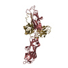
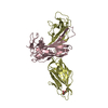
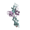
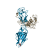
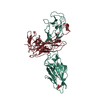
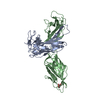
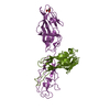
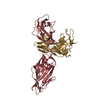
- PDB-1qun: X-RAY STRUCTURE OF THE FIMC-FIMH CHAPERONE ADHESIN COMPLEX FROM U... -
+ Open data

- Basic information
| Entry | Database: PDB / ID: 1qun | ||||||
|---|---|---|---|---|---|---|---|
| Title | X-RAY STRUCTURE OF THE FIMC-FIMH CHAPERONE ADHESIN COMPLEX FROM UROPATHOGENIC E.COLI | ||||||
 Components Components |
| ||||||
 Keywords Keywords | CHAPERONE/STRUCTURAL PROTEIN / CHAPERONE ADHESIN DONOR STRAND COMPLEMENTATION / CHAPERONE-STRUCTURAL PROTEIN COMPLEX | ||||||
| Function / homology |  Function and homology information Function and homology informationpilus tip / mechanosensory behavior / Attachment of bacteria to epithelial cells / cell adhesion involved in single-species biofilm formation / pilus / cell-substrate adhesion / D-mannose binding / host cell membrane / protein folding chaperone / cell wall organization ...pilus tip / mechanosensory behavior / Attachment of bacteria to epithelial cells / cell adhesion involved in single-species biofilm formation / pilus / cell-substrate adhesion / D-mannose binding / host cell membrane / protein folding chaperone / cell wall organization / outer membrane-bounded periplasmic space / protein folding / cell adhesion Similarity search - Function | ||||||
| Biological species |  | ||||||
| Method |  X-RAY DIFFRACTION / SYNCHROTRON / Resolution: 2.8 Å X-RAY DIFFRACTION / SYNCHROTRON / Resolution: 2.8 Å | ||||||
 Authors Authors | Choudhury, D. / Thompson, A. / Stojanoff, V. / Langerman, S. / Pinkner, J. / Hultgren, S.J. / Knight, S. | ||||||
 Citation Citation | Journal: Science / Year: 1999 Title: X-ray structure of the FimC-FimH chaperone-adhesin complex from uropathogenic Escherichia coli. Authors: Choudhury, D. / Thompson, A. / Stojanoff, V. / Langermann, S. / Pinkner, J. / Hultgren, S.J. / Knight, S.D. | ||||||
| History |
|
- Structure visualization
| Structure viewer | Molecule: MolmilJmol/JSmol |
|---|
- Downloads & links
-Download
| PDBx/mmCIF format | 1qun.cif.gz | 712.8 KB | Display | PDBx/mmCIF format |
|---|---|---|---|---|
| PDB format | pdb1qun.ent.gz | 578.8 KB | Display | PDB format |
| PDBx/mmJSON format | 1qun.json.gz | Tree view | PDBx/mmJSON format | |
| Others |  Other downloads Other downloads |
-Validation report
| Arichive directory | https://data.pdbj.org/pub/pdb/validation_reports/qu/1qunftp://data.pdbj.org/pub/pdb/validation_reports/qu/1qun | HTTPS FTP |
|---|
-Related structure data
| Similar structure data |
|---|


-Links
 PDBj
PDBj- Assembly
| Deposited unit | 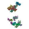
| ||||||||||
|---|---|---|---|---|---|---|---|---|---|---|---|
| 1 | 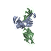
| ||||||||||
| 2 | 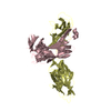
| ||||||||||
| 3 | 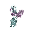
| ||||||||||
| 4 | 
| ||||||||||
| 5 | 
| ||||||||||
| 6 | 
| ||||||||||
| 7 | 
| ||||||||||
| 8 | 
| ||||||||||
| 9 | 
| ||||||||||
| 10 | 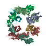
| ||||||||||
| 11 | 
| ||||||||||
| Unit cell |
|
-Components
| #1: Protein | Mass: 22694.023 Da / Num. of mol.: 8 Source method: isolated from a genetically manipulated source Source: (gene. exp.) #2: Protein | Mass: 29081.314 Da / Num. of mol.: 8 Source method: isolated from a genetically manipulated source Source: (gene. exp.) #3: Water | ChemComp-HOH / |  Mass: 18.015 Da / Num. of mol.: 64 / Source method: isolated from a natural source / Formula: H2O Mass: 18.015 Da / Num. of mol.: 64 / Source method: isolated from a natural source / Formula: H2OHas protein modification | Y | |
|---|
-Experimental details
-Experiment
| Experiment | Method: X-RAY DIFFRACTION / Number of used crystals: 1 |
|---|
- Sample preparation
| Crystal | Density Matthews: 2.53 Å3/Da / Density % sol: 51.42 % | ||||||||||||||||||||||||||||||
|---|---|---|---|---|---|---|---|---|---|---|---|---|---|---|---|---|---|---|---|---|---|---|---|---|---|---|---|---|---|---|---|
| Crystal grow | Temperature: 289 K / Method: vapor diffusion, hanging drop / pH: 8.4 Details: Ammonium Sulphate, Tris, pH 8.4, VAPOR DIFFUSION, HANGING DROP, temperature 289K | ||||||||||||||||||||||||||||||
| Crystal grow | *PLUS pH: 8.2 | ||||||||||||||||||||||||||||||
| Components of the solutions | *PLUS
|
-Data collection
| Diffraction | Mean temperature: 173 K |
|---|---|
| Diffraction source | Source: SYNCHROTRON / Site: ESRF  / Beamline: BM14 / Beamline: BM14 |
| Detector | Type: MARRESEARCH / Detector: AREA DETECTOR |
| Radiation | Protocol: SINGLE WAVELENGTH / Monochromatic (M) / Laue (L): M / Scattering type: x-ray |
| Radiation wavelength | Relative weight: 1 |
| Reflection | Resolution: 2.8→20 Å / Num. all: 182983 / Num. obs: 142580 / Observed criterion σ(F): 2 |
| Reflection | *PLUS % possible obs: 98 % / Rmerge(I) obs: 0.076 |
| Reflection shell | *PLUS Rmerge(I) obs: 0.253 |
- Processing
| Software |
| |||||||||||||||||||||||||
|---|---|---|---|---|---|---|---|---|---|---|---|---|---|---|---|---|---|---|---|---|---|---|---|---|---|---|
| Refinement | Resolution: 2.8→20 Å / Cross valid method: THROUGHOUT / σ(F): 2 / Stereochemistry target values: Engh & Huber
| |||||||||||||||||||||||||
| Refinement step | Cycle: LAST / Resolution: 2.8→20 Å
| |||||||||||||||||||||||||
| Refine LS restraints |
| |||||||||||||||||||||||||
| Software | *PLUS Name: REFMAC / Classification: refinement | |||||||||||||||||||||||||
| Refinement | *PLUS Highest resolution: 2.8 Å / σ(F): 2 / % reflection Rfree: 5 % / Rfactor obs: 0.24 / Rfactor Rwork: 0.24 | |||||||||||||||||||||||||
| Solvent computation | *PLUS | |||||||||||||||||||||||||
| Displacement parameters | *PLUS |
