 Movie
Movie Controller

[English] 日本語
 Yorodumi
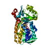
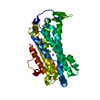
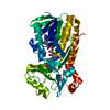
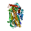
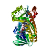
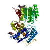
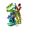
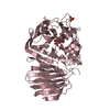
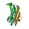
- PDB-2ant: THE 2.6 A STRUCTURE OF ANTITHROMBIN INDICATES A CONFORMATIONAL CH... -
+ Open data

- Basic information
| Entry | Database: PDB / ID: 2ant | ||||||
|---|---|---|---|---|---|---|---|
| Title | THE 2.6 A STRUCTURE OF ANTITHROMBIN INDICATES A CONFORMATIONAL CHANGE AT THE HEPARIN BINDING SITE | ||||||
 Components Components | ANTITHROMBIN | ||||||
 Keywords Keywords | SERPIN / HEPARIN / INHIBITOR | ||||||
| Function / homology |  Function and homology information Function and homology informationregulation of blood coagulation / : / : / Post-translational protein phosphorylation / serine-type endopeptidase inhibitor activity / Regulation of Insulin-like Growth Factor (IGF) transport and uptake by Insulin-like Growth Factor Binding Proteins (IGFBPs) / blood coagulation / heparin binding / extracellular matrix / protease binding ...regulation of blood coagulation / : / : / Post-translational protein phosphorylation / serine-type endopeptidase inhibitor activity / Regulation of Insulin-like Growth Factor (IGF) transport and uptake by Insulin-like Growth Factor Binding Proteins (IGFBPs) / blood coagulation / heparin binding / extracellular matrix / protease binding / blood microparticle / endoplasmic reticulum lumen / : / extracellular exosome / extracellular region / identical protein binding / plasma membrane Similarity search - Function | ||||||
| Biological species |  Homo sapiens (human) Homo sapiens (human) | ||||||
| Method |  X-RAY DIFFRACTION / SYNCHROTRON / MOLECULAR REPLACEMENT / Resolution: 2.6 Å X-RAY DIFFRACTION / SYNCHROTRON / MOLECULAR REPLACEMENT / Resolution: 2.6 Å | ||||||
 Authors Authors | Skinner, R. / Abrahams, J.-P. / Whisstock, J.C. / Lesk, A.M. / Carrell, R.W. / Wardell, M.R. | ||||||
 Citation Citation | Journal: J.Mol.Biol. / Year: 1997 Title: The 2.6 A structure of antithrombin indicates a conformational change at the heparin binding site. Authors: Skinner, R. / Abrahams, J.P. / Whisstock, J.C. / Lesk, A.M. / Carrell, R.W. / Wardell, M.R. #1: Journal: To be PublishedTitle: Improved Diffraction of Antithrombin Crystals Grown in Microgravity Authors: Wardell, M.R. / Skinner, R. / Carter, D.C. / Twigg, P.D. / Abrahams, J.-P. | ||||||
| History |
|
- Structure visualization
| Structure viewer | Molecule: MolmilJmol/JSmol |
|---|
- Downloads & links
-Download
| PDBx/mmCIF format | 2ant.cif.gz | 174.5 KB | Display | PDBx/mmCIF format |
|---|---|---|---|---|
| PDB format | pdb2ant.ent.gz | 139.1 KB | Display | PDB format |
| PDBx/mmJSON format | 2ant.json.gz | Tree view | PDBx/mmJSON format | |
| Others |  Other downloads Other downloads |
-Validation report
| Arichive directory | https://data.pdbj.org/pub/pdb/validation_reports/an/2antftp://data.pdbj.org/pub/pdb/validation_reports/an/2ant | HTTPS FTP |
|---|
-Related structure data
| Related structure data |  1antS S: Starting model for refinement |
|---|---|
| Similar structure data |

-Links
 PDBj
PDBj


- Assembly
| Deposited unit | 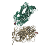
| ||||||||
|---|---|---|---|---|---|---|---|---|---|
| 1 | 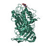
| ||||||||
| 2 | 
| ||||||||
| Unit cell |
|
-Components
| #1: Protein | Mass: 49101.016 Da / Num. of mol.: 2 / Source method: isolated from a natural source Details: ANTITHROMBIN CRYSTALLISED AS A DIMER BETWEEN LATENT MOLECULE AND ONE ACTIVE MOLECULE Source: (natural) Homo sapiens (human) / Tissue: PLASMA / References: UniProt: P01008#2: Sugar | 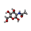  Type: D-saccharide, beta linking / Mass: 221.208 Da / Num. of mol.: 2 Type: D-saccharide, beta linking / Mass: 221.208 Da / Num. of mol.: 2Source method: isolated from a genetically manipulated source Formula: C8H15NO6 #3: Water | ChemComp-HOH / |  Mass: 18.015 Da / Num. of mol.: 79 / Source method: isolated from a natural source / Formula: H2O Mass: 18.015 Da / Num. of mol.: 79 / Source method: isolated from a natural source / Formula: H2OHas protein modification | Y | |
|---|
-Experimental details
-Experiment
| Experiment | Method: X-RAY DIFFRACTION / Number of used crystals: 1 |
|---|
- Sample preparation
| Crystal | Density Matthews: 2.7 Å3/Da / Density % sol: 52.7 % | ||||||||||||||||||||||||||||||||||||
|---|---|---|---|---|---|---|---|---|---|---|---|---|---|---|---|---|---|---|---|---|---|---|---|---|---|---|---|---|---|---|---|---|---|---|---|---|---|
| Crystal grow | pH: 6.7 / Details: 18% PEG 4000 50 MM NA/KPO4 PH 6.7 | ||||||||||||||||||||||||||||||||||||
| Crystal grow | *PLUS Temperature: 4 ℃ / pH: 8 / Method: vapor diffusion, sitting drop | ||||||||||||||||||||||||||||||||||||
| Components of the solutions | *PLUS
|
-Data collection
| Diffraction | Mean temperature: 100 K |
|---|---|
| Diffraction source | Source: SYNCHROTRON / Site: SRS  / Beamline: PX7.2 / Wavelength: 1.488 / Beamline: PX7.2 / Wavelength: 1.488 |
| Detector | Date: Apr 19, 1995 |
| Radiation | Monochromatic (M) / Laue (L): M / Scattering type: x-ray |
| Radiation wavelength | Wavelength: 1.488 Å / Relative weight: 1 |
| Reflection | Resolution: 2.6→30 Å / Num. obs: 24646 / % possible obs: 74 % / Rmerge(I) obs: 0.048 |
| Reflection shell | Resolution: 2.62→2.8 Å / % possible all: 53.9 |
| Reflection | *PLUS Redundancy: 1.8 % |
| Reflection shell | *PLUS % possible obs: 53.9 % |
- Processing
| Software |
| ||||||||||||||||||||||||||||||
|---|---|---|---|---|---|---|---|---|---|---|---|---|---|---|---|---|---|---|---|---|---|---|---|---|---|---|---|---|---|---|---|
| Refinement | Method to determine structure: MOLECULAR REPLACEMENT Starting model: PDB ENTRY 1ANT Resolution: 2.6→26.9 Å / Cross valid method: RANDOM / σ(F): 0
| ||||||||||||||||||||||||||||||
| Refinement step | Cycle: LAST / Resolution: 2.6→26.9 Å
| ||||||||||||||||||||||||||||||
| Refine LS restraints |
| ||||||||||||||||||||||||||||||
| Software | *PLUS Name: TNT / Version: 5D / Classification: refinement | ||||||||||||||||||||||||||||||
| Refinement | *PLUS Rfactor all: 0.217 / Rfactor Rfree: 0.299 | ||||||||||||||||||||||||||||||
| Solvent computation | *PLUS | ||||||||||||||||||||||||||||||
| Displacement parameters | *PLUS | ||||||||||||||||||||||||||||||
| Refine LS restraints | *PLUS
| ||||||||||||||||||||||||||||||
| LS refinement shell | *PLUS Highest resolution: 2.64 Å / Lowest resolution: 2.72 Å / Rfactor Rfree: 0.63 / Rfactor obs: 0.37 |
