 Movie
Movie Controller


+ Open data

- Basic information
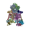
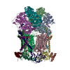
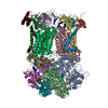
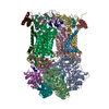
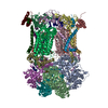
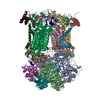
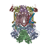
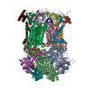
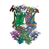
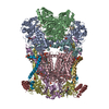
| Entry | Database: PDB / ID: 3h1h | |||||||||
|---|---|---|---|---|---|---|---|---|---|---|
| Title | Cytochrome bc1 complex from chicken | |||||||||
 Components Components |
| |||||||||
 Keywords Keywords | OXIDOREDUCTASE / CYTOCHROME BC1 / MEMBRANE PROTEIN / HEME PROTEIN / RIESKE IRON SULFUR PROTEIN / CYTOCHROME B / CYTOCHROME C1 / COMPLEX III / MITOCHONDRIAL PROCESSING PROTEASE / UBIQUINONE / REDOX ENZYME / RESPIRATORY CHAIN / Electron transport / Heme / Iron / Membrane / Metal-binding / Mitochondrion / Mitochondrion inner membrane / Transmembrane / Transport / Disulfide bond / Iron-sulfur / Transit peptide | |||||||||
| Function / homology |  Function and homology information Function and homology informationMitochondrial translation termination / Respiratory electron transport / Complex III assembly / respiratory chain complex III / quinol-cytochrome-c reductase / quinol-cytochrome-c reductase activity / mitochondrial electron transport, ubiquinol to cytochrome c / respiratory electron transport chain / 2 iron, 2 sulfur cluster binding / response to oxidative stress ...Mitochondrial translation termination / Respiratory electron transport / Complex III assembly / respiratory chain complex III / quinol-cytochrome-c reductase / quinol-cytochrome-c reductase activity / mitochondrial electron transport, ubiquinol to cytochrome c / respiratory electron transport chain / 2 iron, 2 sulfur cluster binding / response to oxidative stress / electron transfer activity / oxidoreductase activity / mitochondrial inner membrane / heme binding / protein-containing complex / mitochondrion / membrane / metal ion binding / cytoplasm Similarity search - Function | |||||||||
| Biological species |  | |||||||||
| Method |  X-RAY DIFFRACTION / SYNCHROTRON / MIR / Resolution: 3.16 Å X-RAY DIFFRACTION / SYNCHROTRON / MIR / Resolution: 3.16 Å | |||||||||
 Authors Authors | Zhang, Z. / Huang, L. / Shulmeister, V.M. / Chi, Y.I. / Kim, K.K. / Hung, L.W. / Crofts, A.R. / Berry, E.A. / Kim, S.H. | |||||||||
 Citation Citation | Journal: Nature / Year: 1998 Title: Electron Transfer by Domain Movement in Cytochrome Bc1 Authors: Zhang, Z. / Huang, L. / Shulmeister, V.M. / Chi, Y.I. / Kim, K.K. / Hung, L.W. / Crofts, A.R. / Berry, E.A. / Kim, S.H. #1: Journal: J.Mol.Biol. / Year: 2005Title: Binding of the Respiratory Chain Inhibitor Antimycin to the Mitochondrial Bc(1) Complex: A New Crystal Structure Reveals an Altered Intramolecular Hydrogen-Bonding Pattern. Authors: Huang, L.S. / Cobessi, D. / Tung, E.Y. / Berry, E.A. #2: Journal: J.BIOENERG.BIOMEMBR. / Year: 1999Title: The Structure of the Avian Mitochondrial Cytochrome bc1 Complex. Authors: Berry, E.A. / Huang, L.S. / Zhang, Z. / Kim, S.H. | |||||||||
| History |
|
- Structure visualization
| Structure viewer | Molecule: MolmilJmol/JSmol |
|---|
- Downloads & links
-Download
| PDBx/mmCIF format | 3h1h.cif.gz | 827.7 KB | Display | PDBx/mmCIF format |
|---|---|---|---|---|
| PDB format | pdb3h1h.ent.gz | 660.7 KB | Display | PDB format |
| PDBx/mmJSON format | 3h1h.json.gz | Tree view | PDBx/mmJSON format | |
| Others |  Other downloads Other downloads |
-Validation report
| Arichive directory | https://data.pdbj.org/pub/pdb/validation_reports/h1/3h1hftp://data.pdbj.org/pub/pdb/validation_reports/h1/3h1h | HTTPS FTP |
|---|
-Related structure data
| Related structure data | 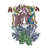 1bccC 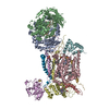 2bccC  3bccC  3h1iC 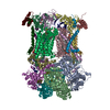 3h1jC  2ppj C: citing same article ( |
|---|---|
| Similar structure data |


-Links
 PDBj
PDBj


- Assembly
| Deposited unit | 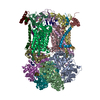
| ||||||||
|---|---|---|---|---|---|---|---|---|---|
| 1 |
| ||||||||
| Unit cell |
|
-Components
-UBIQUINOL-CYTOCHROME-C REDUCTASE COMPLEX CORE PROTEIN ... , 2 types, 4 molecules ANBO
| #1: Protein | Mass: 49503.840 Da / Num. of mol.: 2 / Source method: isolated from a natural source / Source: (natural) References: UniProt: D0VX31*PLUS, quinol-cytochrome-c reductase #2: Protein | Mass: 46683.809 Da / Num. of mol.: 2 / Source method: isolated from a natural source / Source: (natural) |
|---|
-Protein , 2 types, 4 molecules CPDQ
| #3: Protein | Mass: 42622.977 Da / Num. of mol.: 2 / Source method: isolated from a natural source / Source: (natural) #4: Protein | Mass: 26973.744 Da / Num. of mol.: 2 / Source method: isolated from a natural source / Source: (natural) References: UniProt: D0VX26*PLUS, quinol-cytochrome-c reductase |
|---|
-Cytochrome b-c1 complex subunit Rieske, ... , 2 types, 4 molecules ERIV
| #5: Protein | Mass: 21506.188 Da / Num. of mol.: 2 / Fragment: sequence database residues 77-272 / Source method: isolated from a natural source / Source: (natural) #9: Protein/peptide | Mass: 4785.649 Da / Num. of mol.: 2 / Fragment: sequence database residues 1-76 / Source method: isolated from a natural source / Source: (natural) |
|---|
-UBIQUINOL-CYTOCHROME C REDUCTASE COMPLEX ... , 4 types, 8 molecules FSGTHUJW
| #6: Protein | Mass: 13394.397 Da / Num. of mol.: 2 / Source method: isolated from a natural source / Source: (natural) #7: Protein | Mass: 9498.735 Da / Num. of mol.: 2 / Source method: isolated from a natural source / Source: (natural) References: UniProt: D0VX32*PLUS, quinol-cytochrome-c reductase #8: Protein | Mass: 9057.119 Da / Num. of mol.: 2 / Source method: isolated from a natural source / Source: (natural) #10: Protein | Mass: 7005.963 Da / Num. of mol.: 2 / Source method: isolated from a natural source / Source: (natural) References: UniProt: D0VX27*PLUS, quinol-cytochrome-c reductase |
|---|
-Sugars , 1 types, 5 molecules 
| #18: Sugar | ChemComp-BOG /  Type: D-saccharide / Mass: 292.369 Da / Num. of mol.: 5 Type: D-saccharide / Mass: 292.369 Da / Num. of mol.: 5Source method: isolated from a genetically manipulated source Formula: C14H28O6 / Comment: detergent*YM |
|---|
-Non-polymers , 9 types, 50 molecules 
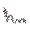


| #11: Chemical | ChemComp-UNL / Num. of mol.: 9 / Source method: obtained synthetically #12: Chemical | ChemComp-HEM /  Mass: 616.487 Da / Num. of mol.: 4 / Source method: obtained synthetically / Formula: C34H32FeN4O4 Mass: 616.487 Da / Num. of mol.: 4 / Source method: obtained synthetically / Formula: C34H32FeN4O4#13: Chemical |  Mass: 863.343 Da / Num. of mol.: 2 / Source method: obtained synthetically / Formula: C59H90O4 Mass: 863.343 Da / Num. of mol.: 2 / Source method: obtained synthetically / Formula: C59H90O4#14: Chemical | ChemComp-CDL /  Mass: 1464.043 Da / Num. of mol.: 4 / Source method: obtained synthetically / Formula: C81H156O17P2 / Comment: phospholipid*YM Mass: 1464.043 Da / Num. of mol.: 4 / Source method: obtained synthetically / Formula: C81H156O17P2 / Comment: phospholipid*YM#15: Chemical | ChemComp-PEE /  Mass: 744.034 Da / Num. of mol.: 6 / Source method: obtained synthetically / Formula: C41H78NO8P / Comment: DOPE, phospholipid*YM Mass: 744.034 Da / Num. of mol.: 6 / Source method: obtained synthetically / Formula: C41H78NO8P / Comment: DOPE, phospholipid*YM#16: Chemical |  Mass: 92.094 Da / Num. of mol.: 2 / Source method: obtained synthetically / Formula: C3H8O3 Mass: 92.094 Da / Num. of mol.: 2 / Source method: obtained synthetically / Formula: C3H8O3#17: Chemical |  Mass: 618.503 Da / Num. of mol.: 2 / Source method: obtained synthetically / Formula: C34H34FeN4O4 Mass: 618.503 Da / Num. of mol.: 2 / Source method: obtained synthetically / Formula: C34H34FeN4O4#19: Chemical |  Mass: 175.820 Da / Num. of mol.: 2 / Source method: obtained synthetically / Formula: Fe2S2 Mass: 175.820 Da / Num. of mol.: 2 / Source method: obtained synthetically / Formula: Fe2S2#20: Water | ChemComp-HOH / | Mass: 18.015 Da / Num. of mol.: 19 / Source method: isolated from a natural source / Formula: H2O |
|---|
-Details
| Has protein modification | Y |
|---|---|
| Sequence details | IN THE COORDINATES THE FIRST 15 RESIDUES IN CHAINS I AND V ARE MODELED AS UNK BECAUSE THE SEQUENCE ...IN THE COORDINATE |
-Experimental details
-Experiment
| Experiment | Method: X-RAY DIFFRACTION / Number of used crystals: 1 |
|---|
- Sample preparation
| Crystal | Density Matthews: 4.03 Å3/Da / Density % sol: 69.47 % |
|---|---|
| Crystal grow | Temperature: 277 K / Method: vapor diffusion, sitting drop / pH: 6.7 Details: 20MM KMES PH 6.7, 75MM NACL, 10% GLYCEROL, AND 6% PEG4000, VAPOR DIFFUSION, SITTING DROP, temperature 277K |
-Data collection
| Diffraction | Mean temperature: 100 K |
|---|---|
| Diffraction source | Source: SYNCHROTRON / Site: SSRL  / Beamline: BL7-1 / Wavelength: 1.08 / Wavelength: 1.08 Å / Beamline: BL7-1 / Wavelength: 1.08 / Wavelength: 1.08 Å |
| Detector | Type: MARRESEARCH / Detector: IMAGE PLATE / Date: Mar 20, 1995 / Details: CYL.-BENT MIRRORS |
| Radiation | Monochromator: SI / Protocol: SINGLE WAVELENGTH / Monochromatic (M) / Laue (L): M / Scattering type: x-ray |
| Radiation wavelength | Wavelength: 1.08 Å / Relative weight: 1 |
| Reflection | Resolution: 3.16→69.338 Å / Num. all: 123869 / Num. obs: 123869 / % possible obs: 91.6 % / Observed criterion σ(I): -2 / Redundancy: 4.5 % / Biso Wilson estimate: 79.4 Å2 / Rmerge(I) obs: 0.095 / Rsym value: 0.102 / Net I/σ(I): 12.5 |
| Reflection shell | Resolution: 3.16→3.27 Å / Redundancy: 3.32 % / Rmerge(I) obs: 0.324 / Mean I/σ(I) obs: 2.7 / Rsym value: 0.4 / % possible all: 84.5 |
- Processing
| Software |
| ||||||||||||||||||||||||||||||||||||
|---|---|---|---|---|---|---|---|---|---|---|---|---|---|---|---|---|---|---|---|---|---|---|---|---|---|---|---|---|---|---|---|---|---|---|---|---|---|
| Refinement | Method to determine structure: MIR / Resolution: 3.16→20 Å / Rfactor Rfree error: 0.006 / Data cutoff high absF: 4383576.42 / Data cutoff low absF: 0 / Isotropic thermal model: RESTRAINED / Cross valid method: THROUGHOUT / σ(F): 0 / Stereochemistry target values: Engh & Huber Details: Heavy Atoms in derivatives of chicken bc1 crystals were located using XtalView, then refined and used for phase calculation in ccp4 mlphare. Nonisomorphous crystals of beef, rabbit bc1 were ...Details: Heavy Atoms in derivatives of chicken bc1 crystals were located using XtalView, then refined and used for phase calculation in ccp4 mlphare. Nonisomorphous crystals of beef, rabbit bc1 were solved by molecular replacement using cut-out density. The phases were improved by cross-crystal and NCS density averaging using RAVE from USF. Model building was carried out in the best native chicken crystal, resulting in structure 1bcc. This structure is a further refinement against the original data, with correct sequences for the subunits and with minor errors in the orifginal tracing corrected.
| ||||||||||||||||||||||||||||||||||||
| Solvent computation | Solvent model: FLAT MODEL / Bsol: 33.7533 Å2 / ksol: 0.271593 e/Å3 | ||||||||||||||||||||||||||||||||||||
| Displacement parameters | Biso mean: 82 Å2
| ||||||||||||||||||||||||||||||||||||
| Refine analyze |
| ||||||||||||||||||||||||||||||||||||
| Refinement step | Cycle: LAST / Resolution: 3.16→20 Å
| ||||||||||||||||||||||||||||||||||||
| Refine LS restraints |
| ||||||||||||||||||||||||||||||||||||
| Refine LS restraints NCS | NCS model details: CONSTR | ||||||||||||||||||||||||||||||||||||
| LS refinement shell | Resolution: 3.16→3.33 Å / Rfactor Rfree error: 0.023 / Total num. of bins used: 7
| ||||||||||||||||||||||||||||||||||||
| Xplor file |
|
