 Movie
Movie Controller

[English] 日本語
 Yorodumi
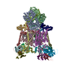
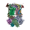
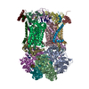
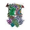
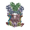
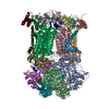
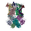
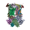
- PDB-3h1k: Chicken cytochrome BC1 complex with ZN++ and an iodinated derivat... -
+ Open data

- Basic information
| Entry | Database: PDB / ID: 3h1k | |||||||||
|---|---|---|---|---|---|---|---|---|---|---|
| Title | Chicken cytochrome BC1 complex with ZN++ and an iodinated derivative of kresoxim-methyl bound | |||||||||
 Components Components |
| |||||||||
 Keywords Keywords | OXIDOREDUCTASE / CYTOCHROME BC1 / MEMBRANE PROTEIN / HEME PROTEIN / RIESKE IRON SULFUR PROTEIN / CYTOCHROME B / CYTOCHROME C1 / COMPLEX III / UBIQUINONE / REDOX ENZYME / ZINC / KRESOXIM-METHYL / RESPIRATORY CHAIN / ELECTRON TRANSPORT / HEME / INNER MEMBRANE IRON / MEMBRANE / METAL-BINDING / MITOCHONDRION / TRANSMEMBRANE / Iron / Mitochondrion inner membrane / Transport / Disulfide bond / Iron-sulfur / Transit peptide | |||||||||
| Function / homology |  Function and homology information Function and homology informationMitochondrial translation termination / Respiratory electron transport / Complex III assembly / respiratory chain complex / respiratory chain complex III / quinol-cytochrome-c reductase / quinol-cytochrome-c reductase activity / mitochondrial electron transport, ubiquinol to cytochrome c / respiratory electron transport chain / 2 iron, 2 sulfur cluster binding ...Mitochondrial translation termination / Respiratory electron transport / Complex III assembly / respiratory chain complex / respiratory chain complex III / quinol-cytochrome-c reductase / quinol-cytochrome-c reductase activity / mitochondrial electron transport, ubiquinol to cytochrome c / respiratory electron transport chain / 2 iron, 2 sulfur cluster binding / response to oxidative stress / electron transfer activity / oxidoreductase activity / mitochondrial inner membrane / heme binding / protein-containing complex / mitochondrion / membrane / metal ion binding / cytoplasm Similarity search - Function | |||||||||
| Biological species |  | |||||||||
| Method |  X-RAY DIFFRACTION / SYNCHROTRON / RIGID BODY REFINEMENT / Resolution: 3.48 Å X-RAY DIFFRACTION / SYNCHROTRON / RIGID BODY REFINEMENT / Resolution: 3.48 Å | |||||||||
 Authors Authors | Berry, E.A. / Zhang, Z. / Bellamy, H.D. / Huang, L.S. | |||||||||
 Citation Citation | Journal: Biochim.Biophys.Acta / Year: 2000 Title: Crystallographic location of two Zn(2+)-binding sites in the avian cytochrome bc(1) complex Authors: Berry, E.A. / Zhang, Z. / Bellamy, H.D. / Huang, L. | |||||||||
| History |
|
- Structure visualization
| Structure viewer | Molecule: MolmilJmol/JSmol |
|---|
- Downloads & links
-Download
| PDBx/mmCIF format | 3h1k.cif.gz | 834.9 KB | Display | PDBx/mmCIF format |
|---|---|---|---|---|
| PDB format | pdb3h1k.ent.gz | 663.4 KB | Display | PDB format |
| PDBx/mmJSON format | 3h1k.json.gz | Tree view | PDBx/mmJSON format | |
| Others |  Other downloads Other downloads |
-Validation report
| Arichive directory | https://data.pdbj.org/pub/pdb/validation_reports/h1/3h1kftp://data.pdbj.org/pub/pdb/validation_reports/h1/3h1k | HTTPS FTP |
|---|
-Related structure data
| Related structure data | 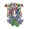 1bccS  2ppj S: Starting model for refinement |
|---|---|
| Similar structure data |


-Links
 PDBj
PDBj


- Assembly
| Deposited unit | 
| ||||||||
|---|---|---|---|---|---|---|---|---|---|
| 1 |
| ||||||||
| Unit cell |
|
-Components
-MITOCHONDRIAL UBIQUINOL-CYTOCHROME-C REDUCTASE COMPLEX CORE PROTEIN ... , 2 types, 4 molecules ANBO
| #1: Protein | Mass: 49503.840 Da / Num. of mol.: 2 / Source method: isolated from a natural source / Source: (natural) References: UniProt: D0VX31*PLUS, quinol-cytochrome-c reductase #2: Protein | Mass: 46683.809 Da / Num. of mol.: 2 / Source method: isolated from a natural source / Source: (natural) References: UniProt: D0VX29*PLUS, quinol-cytochrome-c reductase |
|---|
-Protein , 2 types, 4 molecules CPDQ
| #3: Protein | Mass: 42622.977 Da / Num. of mol.: 2 / Source method: isolated from a natural source / Source: (natural) #4: Protein | Mass: 26973.744 Da / Num. of mol.: 2 / Source method: isolated from a natural source / Source: (natural) References: UniProt: D0VX26*PLUS, quinol-cytochrome-c reductase |
|---|
-Cytochrome b-c1 complex subunit Rieske, ... , 2 types, 4 molecules ERIV
| #5: Protein | Mass: 21506.188 Da / Num. of mol.: 2 / Fragment: sequence database residues 77-272 / Source method: isolated from a natural source / Source: (natural) #9: Protein/peptide | Mass: 4785.649 Da / Num. of mol.: 2 / Fragment: sequence database residues 1-76 / Source method: isolated from a natural source / Source: (natural) |
|---|
-MITOCHONDRIAL UBIQUINOL-CYTOCHROME C REDUCTASE ... , 4 types, 8 molecules FSGTHUJW
| #6: Protein | Mass: 13394.397 Da / Num. of mol.: 2 / Source method: isolated from a natural source / Source: (natural) References: UniProt: D0VX30*PLUS, quinol-cytochrome-c reductase #7: Protein | Mass: 9498.735 Da / Num. of mol.: 2 / Source method: isolated from a natural source / Source: (natural) References: UniProt: D0VX32*PLUS, quinol-cytochrome-c reductase #8: Protein | Mass: 9057.119 Da / Num. of mol.: 2 / Source method: isolated from a natural source / Source: (natural) References: UniProt: D0VX28*PLUS, quinol-cytochrome-c reductase #10: Protein | Mass: 7005.963 Da / Num. of mol.: 2 / Source method: isolated from a natural source / Source: (natural) References: UniProt: D0VX27*PLUS, quinol-cytochrome-c reductase |
|---|
-Sugars , 1 types, 5 molecules 
| #20: Sugar | ChemComp-BOG /  Type: D-saccharide / Mass: 292.369 Da / Num. of mol.: 5 Type: D-saccharide / Mass: 292.369 Da / Num. of mol.: 5Source method: isolated from a genetically manipulated source Formula: C14H28O6 / Comment: detergent*YM |
|---|
-Non-polymers , 11 types, 48 molecules 


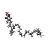


| #11: Chemical | ChemComp-PEE /  Mass: 744.034 Da / Num. of mol.: 6 / Source method: obtained synthetically / Formula: C41H78NO8P / Comment: DOPE, phospholipid*YM Mass: 744.034 Da / Num. of mol.: 6 / Source method: obtained synthetically / Formula: C41H78NO8P / Comment: DOPE, phospholipid*YM#12: Chemical | ChemComp-UNL / Num. of mol.: 5 / Source method: obtained synthetically #13: Chemical | ChemComp-HEM /  Mass: 616.487 Da / Num. of mol.: 4 / Source method: obtained synthetically / Formula: C34H32FeN4O4 Mass: 616.487 Da / Num. of mol.: 4 / Source method: obtained synthetically / Formula: C34H32FeN4O4#14: Chemical |  Mass: 453.271 Da / Num. of mol.: 2 / Source method: obtained synthetically / Formula: C19H20INO4 Mass: 453.271 Da / Num. of mol.: 2 / Source method: obtained synthetically / Formula: C19H20INO4#15: Chemical |  Mass: 863.343 Da / Num. of mol.: 2 / Source method: obtained synthetically / Formula: C59H90O4 Mass: 863.343 Da / Num. of mol.: 2 / Source method: obtained synthetically / Formula: C59H90O4#16: Chemical | ChemComp-CDL /  Mass: 1464.043 Da / Num. of mol.: 4 / Source method: obtained synthetically / Formula: C81H156O17P2 / Comment: phospholipid*YM Mass: 1464.043 Da / Num. of mol.: 4 / Source method: obtained synthetically / Formula: C81H156O17P2 / Comment: phospholipid*YM#17: Chemical |  Mass: 65.409 Da / Num. of mol.: 2 / Source method: obtained synthetically / Formula: Zn Mass: 65.409 Da / Num. of mol.: 2 / Source method: obtained synthetically / Formula: Zn#18: Chemical |  Mass: 92.094 Da / Num. of mol.: 2 / Source method: obtained synthetically / Formula: C3H8O3 Mass: 92.094 Da / Num. of mol.: 2 / Source method: obtained synthetically / Formula: C3H8O3#19: Chemical |  Mass: 618.503 Da / Num. of mol.: 2 / Source method: obtained synthetically / Formula: C34H34FeN4O4 Mass: 618.503 Da / Num. of mol.: 2 / Source method: obtained synthetically / Formula: C34H34FeN4O4#21: Chemical |  Mass: 175.820 Da / Num. of mol.: 2 / Source method: obtained synthetically / Formula: Fe2S2 Mass: 175.820 Da / Num. of mol.: 2 / Source method: obtained synthetically / Formula: Fe2S2#22: Water | ChemComp-HOH / | Mass: 18.015 Da / Num. of mol.: 17 / Source method: isolated from a natural source / Formula: H2O |
|---|
-Details
| Sequence details | IN THE COORDINATES THE FIRST 15 RESIDUES IN CHAINS I AND V ARE MODELED AS UNK BECAUSE THE SEQUENCE ...IN THE COORDINATE |
|---|
-Experimental details
-Experiment
| Experiment | Method: X-RAY DIFFRACTION / Number of used crystals: 1 |
|---|
- Sample preparation
| Crystal | Density Matthews: 4.06 Å3/Da / Density % sol: 69.74 % |
|---|---|
| Crystal grow | Temperature: 277 K / Method: vapor diffusion, sitting drop / pH: 6.7 Details: 20MM KMES PH 6.7, 75MM NACL, 10% GLYCEROL, AND 6% PEG4000. The Kresoxim-methyl derivative WAS ADDED to the protein FROM ETHANOLIC SOLUTION. After verifying good diffraction by these ...Details: 20MM KMES PH 6.7, 75MM NACL, 10% GLYCEROL, AND 6% PEG4000. The Kresoxim-methyl derivative WAS ADDED to the protein FROM ETHANOLIC SOLUTION. After verifying good diffraction by these crystals, some were transferred to a drop of mother liquor supplemented with glycerol and ~0.2 mM ZnCl2. After 1 week this crystal was flash-cooled for data collection. During analysis of Zn binding presence of the inhibitor was overlooked, and in the primary citation publication the anomalous signal of I in the inhibitor was mistakenly attributed to a second Zn binding site, Zn02. VAPOR DIFFUSION, SITTING DROP, temperature 277K |
-Data collection
| Diffraction | Mean temperature: 100 K |
|---|---|
| Diffraction source | Source: SYNCHROTRON / Site: SSRL  / Beamline: BL1-5 / Wavelength: 1.283 / Wavelength: 1.283 Å / Beamline: BL1-5 / Wavelength: 1.283 / Wavelength: 1.283 Å |
| Detector | Type: ADSC QUANTUM 4 / Detector: CCD / Date: Jan 25, 1998 |
| Radiation | Protocol: SINGLE WAVELENGTH / Monochromatic (M) / Laue (L): M / Scattering type: x-ray |
| Radiation wavelength | Wavelength: 1.283 Å / Relative weight: 1 |
| Reflection | Resolution: 3.48→42.61 Å / Num. all: 87072 / Num. obs: 87072 / % possible obs: 89.9 % / Observed criterion σ(I): -3 / Redundancy: 8.5 % / Biso Wilson estimate: 84.8 Å2 / Rsym value: 0.204 / Net I/σ(I): 9.49 |
| Reflection shell | Resolution: 3.48→3.55 Å / Redundancy: 6.8 % / Mean I/σ(I) obs: 1.14 / Num. unique all: 3144 / Rsym value: 0.99 / % possible all: 65.2 |
- Processing
| Software |
| ||||||||||||||||||||||||||||||||||||||||||||||||||||||||||||||||||||||||||||||||
|---|---|---|---|---|---|---|---|---|---|---|---|---|---|---|---|---|---|---|---|---|---|---|---|---|---|---|---|---|---|---|---|---|---|---|---|---|---|---|---|---|---|---|---|---|---|---|---|---|---|---|---|---|---|---|---|---|---|---|---|---|---|---|---|---|---|---|---|---|---|---|---|---|---|---|---|---|---|---|---|---|---|
| Refinement | Method to determine structure: RIGID BODY REFINEMENT Starting model: 1BCC after further correction/refinement Resolution: 3.48→18 Å / Rfactor Rfree error: 0.006 / Data cutoff high absF: 4943137.94 / Data cutoff low absF: 0 / Isotropic thermal model: RESTRAINED / Cross valid method: THROUGHOUT / σ(F): 0 / Stereochemistry target values: Engh & Huber
| ||||||||||||||||||||||||||||||||||||||||||||||||||||||||||||||||||||||||||||||||
| Solvent computation | Solvent model: FLAT MODEL / Bsol: 56.8112 Å2 / ksol: 0.26 e/Å3 | ||||||||||||||||||||||||||||||||||||||||||||||||||||||||||||||||||||||||||||||||
| Displacement parameters | Biso mean: 110.4 Å2
| ||||||||||||||||||||||||||||||||||||||||||||||||||||||||||||||||||||||||||||||||
| Refine analyze |
| ||||||||||||||||||||||||||||||||||||||||||||||||||||||||||||||||||||||||||||||||
| Refinement step | Cycle: LAST / Resolution: 3.48→18 Å
| ||||||||||||||||||||||||||||||||||||||||||||||||||||||||||||||||||||||||||||||||
| Refine LS restraints |
| ||||||||||||||||||||||||||||||||||||||||||||||||||||||||||||||||||||||||||||||||
| Refine LS restraints NCS | NCS model details: CONSTR | ||||||||||||||||||||||||||||||||||||||||||||||||||||||||||||||||||||||||||||||||
| LS refinement shell | Resolution: 3.48→3.66 Å / Rfactor Rfree error: 0.022 / Total num. of bins used: 7
| ||||||||||||||||||||||||||||||||||||||||||||||||||||||||||||||||||||||||||||||||
| Xplor file |
|
