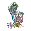
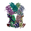
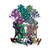
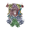
 Movie
Movie Controller


+ Open data

- Basic information
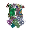
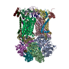
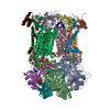
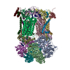
| Entry | Database: PDB / ID: 3l73 | |||||||||
|---|---|---|---|---|---|---|---|---|---|---|
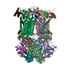
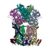
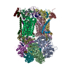
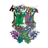
| Title | Cytochrome BC1 complex from chicken with triazolone inhibitor | |||||||||
 Components Components |
| |||||||||
 Keywords Keywords | OXIDOREDUCTASE / CYTOCHROME BC1 / MEMBRANE PROTEIN / HEME PROTEIN / RIESKE IRON SULFUR PROTEIN / CYTOCHROME B / CYTOCHROME C1 / COMPLEX III / MITOCHONDRIAL PROCESSING PROTEIN / UBIQUINONE / AZOXYSTROBIN OXIDOREDUCTASE / REDOX ENZYME RESPIRATORY CHAIN / ELECTRON TRANSPORT / HEME / INNER MEMBRANE / MEMBRANE / STROBILURINS BINDING / MITOCHONDRION / TRANSMEMBRANE / STIGMATELLIN / IRON / MITOCHONDRIAL INNER MEMBRANE / RESPIRATORY CHAIN / IRON-SULFUR / TRANSIT PEPTIDE / Metal-binding / Mitochondrion inner membrane / Transport / Disulfide bond | |||||||||
| Function / homology |  Function and homology information Function and homology informationMitochondrial translation termination / Respiratory electron transport / Complex III assembly / respiratory chain complex / respiratory chain complex III / quinol-cytochrome-c reductase / quinol-cytochrome-c reductase activity / mitochondrial electron transport, ubiquinol to cytochrome c / respiratory electron transport chain / 2 iron, 2 sulfur cluster binding ...Mitochondrial translation termination / Respiratory electron transport / Complex III assembly / respiratory chain complex / respiratory chain complex III / quinol-cytochrome-c reductase / quinol-cytochrome-c reductase activity / mitochondrial electron transport, ubiquinol to cytochrome c / respiratory electron transport chain / 2 iron, 2 sulfur cluster binding / response to oxidative stress / electron transfer activity / oxidoreductase activity / mitochondrial inner membrane / heme binding / protein-containing complex / mitochondrion / membrane / metal ion binding / cytoplasm Similarity search - Function | |||||||||
| Biological species |  | |||||||||
| Method |  X-RAY DIFFRACTION / SYNCHROTRON / RIGID BODY REFINEMENT / Resolution: 3.04 Å X-RAY DIFFRACTION / SYNCHROTRON / RIGID BODY REFINEMENT / Resolution: 3.04 Å | |||||||||
 Authors Authors | Huang, L. / Berry, E.A. | |||||||||
 Citation Citation | Journal: To be Published Title: Famoxadone and related inhibitors bind like methoxy acrylate inhibitors in the Qo site of the BC1 compl and fix the rieske iron-sulfur protein in a positio close to but distinct from that seen ...Title: Famoxadone and related inhibitors bind like methoxy acrylate inhibitors in the Qo site of the BC1 compl and fix the rieske iron-sulfur protein in a positio close to but distinct from that seen with stigmatellin and other "DISTAL" Qo inhibitors. Authors: Huang, L. / Berry, E.A. | |||||||||
| History |
|
- Structure visualization
| Structure viewer | Molecule: MolmilJmol/JSmol |
|---|
- Downloads & links
-Download
| PDBx/mmCIF format | 3l73.cif.gz | 827.9 KB | Display | PDBx/mmCIF format |
|---|---|---|---|---|
| PDB format | pdb3l73.ent.gz | 661 KB | Display | PDB format |
| PDBx/mmJSON format | 3l73.json.gz | Tree view | PDBx/mmJSON format | |
| Others |  Other downloads Other downloads |
-Validation report
| Arichive directory | https://data.pdbj.org/pub/pdb/validation_reports/l7/3l73ftp://data.pdbj.org/pub/pdb/validation_reports/l7/3l73 | HTTPS FTP |
|---|
-Related structure data
| Related structure data |  3l71C  3l72C  3l74C  3h1hS C: citing same article ( S: Starting model for refinement |
|---|---|
| Similar structure data |


-Links
 PDBj
PDBj


- Assembly
| Deposited unit | 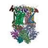
| ||||||||
|---|---|---|---|---|---|---|---|---|---|
| 1 |
| ||||||||
| Unit cell |
| ||||||||
| Details | The deposited coordinates (20 chains plus hetero groups) make up the asymmetric unit which is the biological assembly. One other subunit of the biological assembly (Subunit 11) is lost during purification or crystallization and is not present in the deposited structure. |
-Components
-MITOCHONDRIAL UBIQUINOL-CYTOCHROME-C REDUCTASE COMPLEX CORE PROTEIN ... , 2 types, 4 molecules ANBO
| #1: Protein | Mass: 49503.840 Da / Num. of mol.: 2 / Source method: isolated from a natural source / Source: (natural) #2: Protein | Mass: 46683.809 Da / Num. of mol.: 2 / Source method: isolated from a natural source / Source: (natural) |
|---|
-Protein , 2 types, 4 molecules CPDQ
| #3: Protein | Mass: 42622.977 Da / Num. of mol.: 2 / Source method: isolated from a natural source / Source: (natural) #4: Protein | Mass: 26973.744 Da / Num. of mol.: 2 / Source method: isolated from a natural source / Source: (natural) |
|---|
-CYTOCHROME B-C1 COMPLEX SUBUNIT ... , 2 types, 4 molecules ERIV
| #5: Protein | Mass: 21506.188 Da / Num. of mol.: 2 / Fragment: UNP RESIDUES 77-272 / Source method: isolated from a natural source / Source: (natural) #9: Protein/peptide | Mass: 4785.649 Da / Num. of mol.: 2 / Fragment: UNP RESIDUES 45-76 / Source method: isolated from a natural source / Source: (natural) |
|---|
-MITOCHONDRIAL UBIQUINOL-CYTOCHROME C REDUCTASE ... , 4 types, 8 molecules FSGTHUJW
| #6: Protein | Mass: 13394.397 Da / Num. of mol.: 2 / Source method: isolated from a natural source / Source: (natural) #7: Protein | Mass: 9498.735 Da / Num. of mol.: 2 / Source method: isolated from a natural source / Source: (natural) #8: Protein | Mass: 9057.119 Da / Num. of mol.: 2 / Source method: isolated from a natural source / Source: (natural) #10: Protein | Mass: 7005.963 Da / Num. of mol.: 2 / Source method: isolated from a natural source / Source: (natural) |
|---|
-Sugars , 1 types, 5 molecules 
| #18: Sugar | ChemComp-BOG /  Type: D-saccharide / Mass: 292.369 Da / Num. of mol.: 5 Type: D-saccharide / Mass: 292.369 Da / Num. of mol.: 5Source method: isolated from a genetically manipulated source Formula: C14H28O6 / Comment: detergent*YM |
|---|
-Non-polymers , 9 types, 43 molecules 

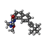
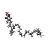


| #11: Chemical | ChemComp-PEE /  Mass: 744.034 Da / Num. of mol.: 6 / Source method: obtained synthetically / Formula: C41H78NO8P / Comment: DOPE, phospholipid*YM Mass: 744.034 Da / Num. of mol.: 6 / Source method: obtained synthetically / Formula: C41H78NO8P / Comment: DOPE, phospholipid*YM#12: Chemical | ChemComp-HEM /  Mass: 616.487 Da / Num. of mol.: 4 / Source method: obtained synthetically / Formula: C34H32FeN4O4 Mass: 616.487 Da / Num. of mol.: 4 / Source method: obtained synthetically / Formula: C34H32FeN4O4#13: Chemical |  Mass: 335.400 Da / Num. of mol.: 2 / Source method: obtained synthetically / Formula: C20H21N3O2 Mass: 335.400 Da / Num. of mol.: 2 / Source method: obtained synthetically / Formula: C20H21N3O2#14: Chemical |  Mass: 863.343 Da / Num. of mol.: 2 / Source method: obtained synthetically / Formula: C59H90O4 Mass: 863.343 Da / Num. of mol.: 2 / Source method: obtained synthetically / Formula: C59H90O4#15: Chemical |  Mass: 92.094 Da / Num. of mol.: 2 / Source method: obtained synthetically / Formula: C3H8O3 Mass: 92.094 Da / Num. of mol.: 2 / Source method: obtained synthetically / Formula: C3H8O3#16: Chemical |  Mass: 618.503 Da / Num. of mol.: 2 / Source method: obtained synthetically / Formula: C34H34FeN4O4 Mass: 618.503 Da / Num. of mol.: 2 / Source method: obtained synthetically / Formula: C34H34FeN4O4#17: Chemical | ChemComp-CDL /  Mass: 1464.043 Da / Num. of mol.: 4 / Source method: obtained synthetically / Formula: C81H156O17P2 / Comment: phospholipid*YM Mass: 1464.043 Da / Num. of mol.: 4 / Source method: obtained synthetically / Formula: C81H156O17P2 / Comment: phospholipid*YM#19: Chemical |  Mass: 175.820 Da / Num. of mol.: 2 / Source method: obtained synthetically / Formula: Fe2S2 Mass: 175.820 Da / Num. of mol.: 2 / Source method: obtained synthetically / Formula: Fe2S2#20: Water | ChemComp-HOH / | Mass: 18.015 Da / Num. of mol.: 19 / Source method: isolated from a natural source / Formula: H2O |
|---|
-Details
| Sequence details | THE COMPLETE SEQUENCE OF CHAIN I AND V IS ...THE COMPLETE SEQUENCE OF CHAIN I AND V IS MLSVAARSGP |
|---|
-Experimental details
-Experiment
| Experiment | Method: X-RAY DIFFRACTION / Number of used crystals: 1 |
|---|
- Sample preparation
| Crystal | Density Matthews: 4.02 Å3/Da / Density % sol: 69.38 % |
|---|---|
| Crystal grow | Temperature: 277 K / Method: vapor diffusion, sitting drop / pH: 6.77 Details: FINAL CONCENTRATIONS BEFORE DIFFUSION: 50 MM CACODYLATE, 9.4 MM TRISHCL, 10 MM MGCL2, 50 G/L GLYCEROL, 30 G/L PEG 3350DA, 0.23 MM EDTA, 0.47 G/L UNDECYL MALTOSIDE, 31 MM OCTYL GLUCOSIDE, PH ...Details: FINAL CONCENTRATIONS BEFORE DIFFUSION: 50 MM CACODYLATE, 9.4 MM TRISHCL, 10 MM MGCL2, 50 G/L GLYCEROL, 30 G/L PEG 3350DA, 0.23 MM EDTA, 0.47 G/L UNDECYL MALTOSIDE, 31 MM OCTYL GLUCOSIDE, PH 6.77, VAPOR DIFFUSION, SITTING DROP, temperature 277K |
-Data collection
| Diffraction | Mean temperature: 100 K | |||||||||
|---|---|---|---|---|---|---|---|---|---|---|
| Diffraction source | Source: SYNCHROTRON / Site: ALS  / Beamline: 5.0.2 / Wavelength: 1 / Wavelength: 1 Å / Beamline: 5.0.2 / Wavelength: 1 / Wavelength: 1 Å | |||||||||
| Detector | Type: ADSC QUANTUM 315 / Detector: CCD / Date: Aug 26, 2006 | |||||||||
| Radiation | Monochromator: DOUBLE CRYSTAL SI(111) / Protocol: SINGLE WAVELENGTH / Monochromatic (M) / Laue (L): M / Scattering type: x-ray | |||||||||
| Radiation wavelength |
| |||||||||
| Reflection | Resolution: 2.9→25 Å / Num. all: 167200 / Num. obs: 167200 / % possible obs: 99.9 % / Observed criterion σ(I): -3 / Redundancy: 4.3 % / Biso Wilson estimate: 80.8 Å2 / Rsym value: 0.085 / Net I/σ(I): 19.38 | |||||||||
| Reflection shell | Resolution: 2.9→2.95 Å / Redundancy: 3.9 % / Mean I/σ(I) obs: 1.072 / Num. unique all: 8264 / % possible all: 99.7 |
- Processing
| Software |
| ||||||||||||||||||||||||||||||||||||||||||||||||||||||||||||||||||||||||||||||||
|---|---|---|---|---|---|---|---|---|---|---|---|---|---|---|---|---|---|---|---|---|---|---|---|---|---|---|---|---|---|---|---|---|---|---|---|---|---|---|---|---|---|---|---|---|---|---|---|---|---|---|---|---|---|---|---|---|---|---|---|---|---|---|---|---|---|---|---|---|---|---|---|---|---|---|---|---|---|---|---|---|---|
| Refinement | Method to determine structure: RIGID BODY REFINEMENT Starting model: PDB ENTRY 3H1H Resolution: 3.04→24.97 Å / Rfactor Rfree error: 0.005 / Data cutoff high absF: 3513358.57 / Data cutoff low absF: 0 / Isotropic thermal model: RESTRAINED / Cross valid method: THROUGHOUT / σ(F): 0 / Stereochemistry target values: Engh & Huber
| ||||||||||||||||||||||||||||||||||||||||||||||||||||||||||||||||||||||||||||||||
| Solvent computation | Solvent model: FLAT MODEL / Bsol: 21.5201 Å2 / ksol: 0.241878 e/Å3 | ||||||||||||||||||||||||||||||||||||||||||||||||||||||||||||||||||||||||||||||||
| Displacement parameters | Biso mean: 86.5 Å2
| ||||||||||||||||||||||||||||||||||||||||||||||||||||||||||||||||||||||||||||||||
| Refine analyze |
| ||||||||||||||||||||||||||||||||||||||||||||||||||||||||||||||||||||||||||||||||
| Refinement step | Cycle: LAST / Resolution: 3.04→24.97 Å
| ||||||||||||||||||||||||||||||||||||||||||||||||||||||||||||||||||||||||||||||||
| Refine LS restraints |
| ||||||||||||||||||||||||||||||||||||||||||||||||||||||||||||||||||||||||||||||||
| Refine LS restraints NCS | NCS model details: CONSTR | ||||||||||||||||||||||||||||||||||||||||||||||||||||||||||||||||||||||||||||||||
| LS refinement shell | Resolution: 3.04→3.2 Å / Rfactor Rfree error: 0.022 / Total num. of bins used: 7
| ||||||||||||||||||||||||||||||||||||||||||||||||||||||||||||||||||||||||||||||||
| Xplor file |
|
