 Movie
Movie Controller


+ Open data

- Basic information
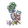
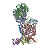
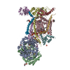
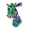
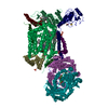
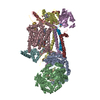
| Entry | Database: PDB / ID: 2bcc | |||||||||
|---|---|---|---|---|---|---|---|---|---|---|
| Title | STIGMATELLIN-BOUND CYTOCHROME BC1 COMPLEX FROM CHICKEN | |||||||||
 Components Components | (UBIQUINOL CYTOCHROME C ...) x 10 | |||||||||
 Keywords Keywords | OXIDOREDUCTASE / UBIQUINONE / REDOX ENZYME / MEMBRANE PROTEIN / RESPIRATORY CHAIN / STIGMATELLIN | |||||||||
| Function / homology |  Function and homology information Function and homology informationMitochondrial translation termination / Respiratory electron transport / Complex III assembly / Complex III assembly / subthalamus development / pons development / cerebellar Purkinje cell layer development / Respiratory electron transport / pyramidal neuron development / thalamus development ...Mitochondrial translation termination / Respiratory electron transport / Complex III assembly / Complex III assembly / subthalamus development / pons development / cerebellar Purkinje cell layer development / Respiratory electron transport / pyramidal neuron development / thalamus development / respiratory chain complex III / quinol-cytochrome-c reductase / quinol-cytochrome-c reductase activity / mitochondrial electron transport, ubiquinol to cytochrome c / Mitochondrial protein degradation / hypothalamus development / midbrain development / respiratory electron transport chain / hippocampus development / metalloendopeptidase activity / 2 iron, 2 sulfur cluster binding / response to oxidative stress / oxidoreductase activity / mitochondrial inner membrane / heme binding / protein-containing complex / mitochondrion / proteolysis / membrane / metal ion binding / cytoplasm Similarity search - Function | |||||||||
| Biological species |  | |||||||||
| Method |  X-RAY DIFFRACTION / SYNCHROTRON / MOLECULAR REPLACEMENT USING NATIVE STRUCTURE SOLVED BY THE SAME AUTHOR / Resolution: 3.5 Å X-RAY DIFFRACTION / SYNCHROTRON / MOLECULAR REPLACEMENT USING NATIVE STRUCTURE SOLVED BY THE SAME AUTHOR / Resolution: 3.5 Å | |||||||||
 Authors Authors | Zhang, Z. / Huang, L. / Shulmeister, V.M. / Chi, Y.I. / Kim, K.K. / Hung, L.W. / Crofts, A.R. / Berry, E.A. / Kim, S.H. | |||||||||
 Citation Citation | Journal: Nature / Year: 1998 Title: Electron Transfer by Domain Movement in Cytochrome Bc1 Authors: Zhang, Z. / Huang, L. / Shulmeister, V.M. / Chi, Y.I. / Kim, K.K. / Hung, L.W. / Crofts, A.R. / Berry, E.A. / Kim, S.H. #1: Journal: Proc.Natl.Acad.Sci.USA / Year: 1999 Title: Pathways for proton release during ubihydroquinone oxidation by the bc(1) complex. Authors: Crofts, A.R. / Hong, S. / Ugulava, N. / Barquera, B. / Gennis, R. / Guergova-Kuras, M. / Berry, E.A. | |||||||||
| History |
| |||||||||
| Remark 300 | BIOMOLECULE DETERGENT SOLUBILIZED ENZYME IS DIMERIC, SAME DIMERIC RELATIONSHIP IS FOUND IN ... BIOMOLECULE DETERGENT SOLUBILIZED ENZYME IS DIMERIC, SAME DIMERIC RELATIONSHIP IS FOUND IN DIFFERENT CRYSTAL FORMS, SOMETIMES ON CRYSTALLOGRAPHIC TWO-FOLD. THIS DIMERIC BIOMOLECULES IS PRESENT IN THE ASYMMETRIC UNIT, HOWEVER THIS PDB ENTRY DOES NOT CONTAIN THE ASYMMETRIC UNIT BUT ONLY ONE MONOMER. SEE REMARK 350 FOR INFORMATION ON GENERATING THE BIOLOGICAL MOLECULE. | |||||||||
| Remark 350 | GENERATING THE BIOMOLECULE COORDINATES FOR A COMPLETE MULTIMER REPRESENTING THE KNOWN BIOLOGICALLY ... GENERATING THE BIOMOLECULE COORDINATES FOR A COMPLETE MULTIMER REPRESENTING THE KNOWN BIOLOGICALLY SIGNIFICANT OLIGOMERIZATION STATE OF THE MOLECULE CAN BE GENERATED BY APPLYING BIOMT TRANSFORMATIONS GIVEN BELOW. BOTH NON-CRYSTALLOGRAPHIC AND CRYSTALLOGRAPHIC OPERATIONS ARE GIVEN. BIOMOLECULE: 1 APPLY THE FOLLOWING TO CHAINS: A, B, C, D, E, F, G, H, I, J BIOMT1 1 -0.836106 -0.548567 -0.000520 130.10969 BIOMT2 1 -0.548555 0.836080 0.007595 38.18410 BIOMT3 1 -0.003732 0.006636 -0.999971 169.17245 |
- Structure visualization
| Structure viewer | Molecule: MolmilJmol/JSmol |
|---|
- Downloads & links
-Download
| PDBx/mmCIF format | 2bcc.cif.gz | 414.5 KB | Display | PDBx/mmCIF format |
|---|---|---|---|---|
| PDB format | pdb2bcc.ent.gz | 324.7 KB | Display | PDB format |
| PDBx/mmJSON format | 2bcc.json.gz | Tree view | PDBx/mmJSON format | |
| Others |  Other downloads Other downloads |
-Validation report
| Arichive directory | https://data.pdbj.org/pub/pdb/validation_reports/bc/2bccftp://data.pdbj.org/pub/pdb/validation_reports/bc/2bcc | HTTPS FTP |
|---|
-Related structure data
| Related structure data | 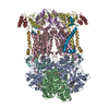 1bccSC  3bccC 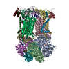 3h1hC 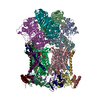 3h1iC 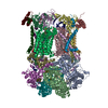 3h1jC S: Starting model for refinement C: citing same article ( |
|---|---|
| Similar structure data |


-Links
 PDBj
PDBj


- Assembly
| Deposited unit | 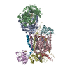
| ||||||||
|---|---|---|---|---|---|---|---|---|---|
| 1 |
| ||||||||
| Unit cell |
| ||||||||
| Noncrystallographic symmetry (NCS) | NCS oper: (Code: generate Matrix: (-0.836106, -0.548567, -0.00052), Vector: |
-Components
-UBIQUINOL CYTOCHROME C ... , 10 types, 10 molecules ABCDEFGHIJ
| #1: Protein | Mass: 49795.859 Da / Num. of mol.: 1 / Source method: isolated from a natural source / Details: ISOLATED FROM TISSUE / Source: (natural) References: UniProt: P31800*PLUS, quinol-cytochrome-c reductase |
|---|---|
| #2: Protein | Mass: 45091.727 Da / Num. of mol.: 1 / Source method: isolated from a natural source / Details: ISOLATED FROM TISSUE / Source: (natural) References: UniProt: P23004*PLUS, quinol-cytochrome-c reductase |
| #3: Protein | Mass: 42622.977 Da / Num. of mol.: 1 / Source method: isolated from a natural source / Details: ISOLATED FROM TISSUE / Source: (natural) |
| #4: Protein | Mass: 27357.250 Da / Num. of mol.: 1 / Source method: isolated from a natural source / Details: ISOLATED FROM TISSUE / Source: (natural) References: UniProt: P00125*PLUS, quinol-cytochrome-c reductase |
| #5: Protein | Mass: 21739.674 Da / Num. of mol.: 1 / Source method: isolated from a natural source / Details: ISOLATED FROM TISSUE / Source: (natural) References: UniProt: P13272*PLUS, quinol-cytochrome-c reductase |
| #6: Protein | Mass: 13400.142 Da / Num. of mol.: 1 / Source method: isolated from a natural source / Details: ISOLATED FROM TISSUE / Source: (natural) References: UniProt: P00129*PLUS, quinol-cytochrome-c reductase |
| #7: Protein | Mass: 9715.241 Da / Num. of mol.: 1 / Source method: isolated from a natural source / Details: ISOLATED FROM TISSUE / Source: (natural) References: UniProt: P13271*PLUS, quinol-cytochrome-c reductase |
| #8: Protein | Mass: 9223.133 Da / Num. of mol.: 1 / Source method: isolated from a natural source / Details: ISOLATED FROM TISSUE / Source: (natural) References: UniProt: P00126*PLUS, quinol-cytochrome-c reductase |
| #9: Protein/peptide | Mass: 2826.475 Da / Num. of mol.: 1 / Source method: isolated from a natural source / Details: ISOLATED FROM TISSUE / Source: (natural) |
| #10: Protein | Mass: 7175.295 Da / Num. of mol.: 1 / Source method: isolated from a natural source / Details: ISOLATED FROM TISSUE / Source: (natural) References: UniProt: P00130*PLUS, quinol-cytochrome-c reductase |
-Sugars , 1 types, 1 molecules 
| #15: Sugar | ChemComp-BOG /  Type: D-saccharide / Mass: 292.369 Da / Num. of mol.: 1 Type: D-saccharide / Mass: 292.369 Da / Num. of mol.: 1Source method: isolated from a genetically manipulated source Formula: C14H28O6 / Comment: detergent*YM |
|---|
-Non-polymers , 5 types, 8 molecules 


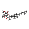

| #11: Chemical |  Mass: 616.487 Da / Num. of mol.: 3 / Source method: obtained synthetically / Formula: C34H32FeN4O4 Mass: 616.487 Da / Num. of mol.: 3 / Source method: obtained synthetically / Formula: C34H32FeN4O4#12: Chemical | ChemComp-U10 / |  Mass: 863.343 Da / Num. of mol.: 1 / Source method: obtained synthetically / Formula: C59H90O4 Mass: 863.343 Da / Num. of mol.: 1 / Source method: obtained synthetically / Formula: C59H90O4#13: Chemical |  Mass: 744.034 Da / Num. of mol.: 2 / Source method: obtained synthetically / Formula: C41H78NO8P / Comment: DOPE, phospholipid*YM Mass: 744.034 Da / Num. of mol.: 2 / Source method: obtained synthetically / Formula: C41H78NO8P / Comment: DOPE, phospholipid*YM#14: Chemical | ChemComp-SIG / |  Mass: 482.651 Da / Num. of mol.: 1 / Source method: obtained synthetically / Formula: C30H42O5 Mass: 482.651 Da / Num. of mol.: 1 / Source method: obtained synthetically / Formula: C30H42O5#16: Chemical | ChemComp-FES / |  Mass: 175.820 Da / Num. of mol.: 1 / Source method: obtained synthetically / Formula: Fe2S2 Mass: 175.820 Da / Num. of mol.: 1 / Source method: obtained synthetically / Formula: Fe2S2 |
|---|
-Details
| Sequence details | THOUGH PROTEINS IN THIS ENTRY ARE FROM CHICKEN, BOVINE SEQUENCES WERE USED FOR MODELING. |
|---|
-Experimental details
-Experiment
| Experiment | Method: X-RAY DIFFRACTION / Number of used crystals: 1 |
|---|
- Sample preparation
| Crystal | Density Matthews: 3.93 Å3/Da / Density % sol: 68.7 % |
|---|---|
| Crystal grow | pH: 6.7 Details: 20MM KMES PH6.7, 75MM NACL, 10% GLYCEROL, AND 6% PEG4000, INHIBITOR WAS ADDED FROM ETHANOLIC SOLUTION |
-Data collection
| Diffraction | Mean temperature: 100 K |
|---|---|
| Diffraction source | Source: SYNCHROTRON / Site: SSRL  / Beamline: BL7-1 / Wavelength: 1.08 / Beamline: BL7-1 / Wavelength: 1.08 |
| Detector | Type: MARRESEARCH / Detector: IMAGE PLATE / Date: Oct 4, 1997 / Details: MIRROR |
| Radiation | Monochromator: SI(111) / Protocol: SINGLE WAVELENGTH / Monochromatic (M) / Laue (L): M / Scattering type: x-ray |
| Radiation wavelength | Wavelength: 1.08 Å / Relative weight: 1 |
| Reflection | Resolution: 3→10000 Å / Num. obs: 117928 / % possible obs: 77.1 % / Redundancy: 2.9 % / Biso Wilson estimate: 19 Å2 / Rmerge(I) obs: 0.131 / Rsym value: 0.131 / Net I/σ(I): 6 |
| Reflection shell | Resolution: 3.38→3.56 Å / Redundancy: 2.2 % / Rmerge(I) obs: 0.459 / Mean I/σ(I) obs: 1.6 / Rsym value: 0.459 / % possible all: 75 |
- Processing
| Software |
| ||||||||||||||||||||||||||||||||||||||||||||||||||||||||||||
|---|---|---|---|---|---|---|---|---|---|---|---|---|---|---|---|---|---|---|---|---|---|---|---|---|---|---|---|---|---|---|---|---|---|---|---|---|---|---|---|---|---|---|---|---|---|---|---|---|---|---|---|---|---|---|---|---|---|---|---|---|---|
| Refinement | Method to determine structure: MOLECULAR REPLACEMENT USING NATIVE STRUCTURE SOLVED BY THE SAME AUTHOR Starting model: PDB 1BCC Resolution: 3.5→12 Å / Rfactor Rfree error: 0.005 / Isotropic thermal model: RESTRAINED / Cross valid method: THROUGHOUT / Details: BULK SOLVENT MODEL USED
| ||||||||||||||||||||||||||||||||||||||||||||||||||||||||||||
| Displacement parameters | Biso mean: 4.6 Å2
| ||||||||||||||||||||||||||||||||||||||||||||||||||||||||||||
| Refine analyze |
| ||||||||||||||||||||||||||||||||||||||||||||||||||||||||||||
| Refinement step | Cycle: LAST / Resolution: 3.5→12 Å
| ||||||||||||||||||||||||||||||||||||||||||||||||||||||||||||
| Refine LS restraints |
| ||||||||||||||||||||||||||||||||||||||||||||||||||||||||||||
| Refine LS restraints NCS | NCS model details: CONSTR | ||||||||||||||||||||||||||||||||||||||||||||||||||||||||||||
| LS refinement shell | Resolution: 3.5→3.71 Å / Rfactor Rfree error: 0.014 / Total num. of bins used: 6
|
