 Movie
Movie Controller


+ Open data

- Basic information
| Entry | Database: PDB / ID: 3g91 | ||||||
|---|---|---|---|---|---|---|---|
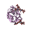
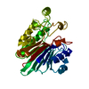
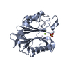
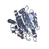
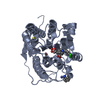
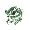
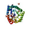
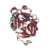
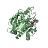
| Title | 1.2 Angstrom structure of the exonuclease III homologue Mth0212 | ||||||
 Components Components | Exodeoxyribonuclease | ||||||
 Keywords Keywords | HYDROLASE / double-strand specific 3'-5' exonuclease / AP endonuclease / 2'-desoxyuridine endonuclease | ||||||
| Function / homology |  Function and homology information Function and homology informationexodeoxyribonuclease III / double-stranded DNA 3'-5' DNA exonuclease activity / phosphoric diester hydrolase activity / class I DNA-(apurinic or apyrimidinic site) endonuclease activity / DNA-(apurinic or apyrimidinic site) lyase / base-excision repair / DNA binding / metal ion binding Similarity search - Function | ||||||
| Biological species |   Methanothermobacter thermautotrophicus (archaea) Methanothermobacter thermautotrophicus (archaea) | ||||||
| Method |  X-RAY DIFFRACTION / SYNCHROTRON / MOLECULAR REPLACEMENT / molecular replacement / Resolution: 1.23 Å X-RAY DIFFRACTION / SYNCHROTRON / MOLECULAR REPLACEMENT / molecular replacement / Resolution: 1.23 Å | ||||||
 Authors Authors | Lakomek, K. / Dickmanns, A. / Ficner, R. | ||||||
 Citation Citation | Journal: J.Mol.Biol. / Year: 2010 Title: Crystal Structure Analysis of DNA Uridine Endonuclease Mth212 Bound to DNA. Authors: Lakomek, K. / Dickmanns, A. / Ciirdaeva, E. / Schomacher, L. / Ficner, R. #1: Journal: Nucleic Acids Res. / Year: 2006 Title: The Methanothermobacter thermautotrophicus ExoIII homologue Mth212 is a DNA uridine endonuclease Authors: Georg, J. / Schomacher, L. / Chong, J.P.J. / Majernik, A.I. / Raabe, M. / Urlaub, H. / Muller, S. / Ciirdaeva, E. / Kramer, W. / Fritz, H.-J. | ||||||
| History |
|
- Structure visualization
| Structure viewer | Molecule: MolmilJmol/JSmol |
|---|
- Downloads & links
-Download
| PDBx/mmCIF format | 3g91.cif.gz | 152.3 KB | Display | PDBx/mmCIF format |
|---|---|---|---|---|
| PDB format | pdb3g91.ent.gz | 117.2 KB | Display | PDB format |
| PDBx/mmJSON format | 3g91.json.gz | Tree view | PDBx/mmJSON format | |
| Others |  Other downloads Other downloads |
-Validation report
| Arichive directory | https://data.pdbj.org/pub/pdb/validation_reports/g9/3g91ftp://data.pdbj.org/pub/pdb/validation_reports/g9/3g91 | HTTPS FTP |
|---|
-Related structure data
| Related structure data |  3fziSC  3g00C  3g0aC  3g0rC  3g1kC  3g2cC  3g2dC  3g38C  3g3cC  3g3yC  3g4tC  3g8vC  3ga6C S: Starting model for refinement C: citing same article ( |
|---|---|
| Similar structure data |

-Links
 PDBj
PDBj


- Assembly
| Deposited unit | 
| ||||||||
|---|---|---|---|---|---|---|---|---|---|
| 1 |
| ||||||||
| Unit cell |
|
-Components
-Protein , 1 types, 1 molecules A
| #1: Protein | Mass: 31372.549 Da / Num. of mol.: 1 / Mutation: T2A, K116A Source method: isolated from a genetically manipulated source Details: strain used for expression lacks the gene ung coding for a uracil DNA glycosylase Source: (gene. exp.) Methanothermobacter thermautotrophicus (archaea)Strain: Delta H (DSM 1053) / Gene: mth0212, MTH212, MTH_212 / Plasmid: pET_B_001-mth212 (K116A) / Production host:  |
|---|
-Non-polymers , 6 types, 438 molecules 


| #2: Chemical | ChemComp-PO4 /  Mass: 94.971 Da / Num. of mol.: 1 / Source method: obtained synthetically / Formula: PO4 Mass: 94.971 Da / Num. of mol.: 1 / Source method: obtained synthetically / Formula: PO4 |
|---|---|
| #3: Chemical | ChemComp-MG /  Mass: 24.305 Da / Num. of mol.: 1 / Source method: obtained synthetically / Formula: Mg Mass: 24.305 Da / Num. of mol.: 1 / Source method: obtained synthetically / Formula: Mg |
| #4: Chemical | ChemComp-GOL /  Mass: 92.094 Da / Num. of mol.: 1 / Source method: obtained synthetically / Formula: C3H8O3 Mass: 92.094 Da / Num. of mol.: 1 / Source method: obtained synthetically / Formula: C3H8O3 |
| #5: Chemical | ChemComp-PEG /  Mass: 106.120 Da / Num. of mol.: 1 / Source method: obtained synthetically / Formula: C4H10O3 Mass: 106.120 Da / Num. of mol.: 1 / Source method: obtained synthetically / Formula: C4H10O3 |
| #6: Chemical | ChemComp-PG4 /  Mass: 194.226 Da / Num. of mol.: 1 / Source method: obtained synthetically / Formula: C8H18O5 / Comment: precipitant*YM Mass: 194.226 Da / Num. of mol.: 1 / Source method: obtained synthetically / Formula: C8H18O5 / Comment: precipitant*YM |
| #7: Water | ChemComp-HOH / Mass: 18.015 Da / Num. of mol.: 433 / Source method: isolated from a natural source / Formula: H2O |
-Experimental details
-Experiment
| Experiment | Method: X-RAY DIFFRACTION / Number of used crystals: 1 |
|---|
- Sample preparation
| Crystal | Density Matthews: 2.08 Å3/Da / Density % sol: 41 % |
|---|---|
| Crystal grow | Temperature: 293 K / Method: vapor diffusion, sitting drop / pH: 6 Details: reservoir solution: 20% (v/v) MPD, 40mM MgAc, 50mM sodium cacodylate pH 6.0; protein solution: 180mM KCl, 10mM KH2PO4/K2HPO4 pH 7.0, VAPOR DIFFUSION, SITTING DROP, temperature 293K |
-Data collection
| Diffraction | Mean temperature: 100 K |
|---|---|
| Diffraction source | Source: SYNCHROTRON / Site: BESSY  / Beamline: 14.2 / Wavelength: 0.91841 Å / Beamline: 14.2 / Wavelength: 0.91841 Å |
| Detector | Type: MARMOSAIC 225 mm CCD / Detector: CCD / Date: Aug 15, 2008 |
| Radiation | Protocol: SINGLE WAVELENGTH / Monochromatic (M) / Laue (L): M / Scattering type: x-ray |
| Radiation wavelength | Wavelength: 0.91841 Å / Relative weight: 1 |
| Reflection | Resolution: 1.23→18 Å / Num. obs: 73200 / % possible obs: 97.9 % / Observed criterion σ(I): -3 / Redundancy: 4.1 % / Biso Wilson estimate: 15.044 Å2 / Rmerge(I) obs: 0.029 / Net I/σ(I): 28.1 |
| Reflection shell | Resolution: 1.23→1.27 Å / Redundancy: 3.6 % / Rmerge(I) obs: 0.131 / Mean I/σ(I) obs: 9 / Num. measured obs: 23038 / Num. unique all: 6790 / Num. unique obs: 6297 / % possible all: 92.7 |
-Phasing
| Phasing | Method: molecular replacement |
|---|
- Processing
| Software |
| |||||||||||||||||||||||||||||||||
|---|---|---|---|---|---|---|---|---|---|---|---|---|---|---|---|---|---|---|---|---|---|---|---|---|---|---|---|---|---|---|---|---|---|---|
| Refinement | Method to determine structure: MOLECULAR REPLACEMENT Starting model: PDB ENTRY 3FZI Resolution: 1.23→9.99 Å / Num. parameters: 24979 / Num. restraintsaints: 32073 / Occupancy max: 1 / Occupancy min: 0 / Cross valid method: THROUGHOUT / Stereochemistry target values: ENGH AND HUBER / Details: anisotropic factor was used in refinement
| |||||||||||||||||||||||||||||||||
| Displacement parameters | Biso max: 153.29 Å2 / Biso mean: 20.046 Å2 / Biso min: 3.27 Å2 | |||||||||||||||||||||||||||||||||
| Refine analyze | Num. disordered residues: 29 / Occupancy sum hydrogen: 0 / Occupancy sum non hydrogen: 2606.71 | |||||||||||||||||||||||||||||||||
| Refinement step | Cycle: LAST / Resolution: 1.23→9.99 Å
| |||||||||||||||||||||||||||||||||
| Refine LS restraints |
|
