 Movie
Movie Controller


+ Open data

- Basic information
| Entry | Database: PDB / ID: 3g2c | ||||||
|---|---|---|---|---|---|---|---|
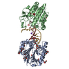
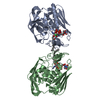
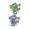
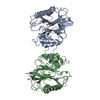
| Title | Mth0212 in complex with a short ssDNA (CGTA) | ||||||
 Components Components |
| ||||||
 Keywords Keywords | HYDROLASE/DNA / protein-DNA complex / single-stranded DNA / flipped nucleotide / PO4 / Mg2+ / HYDROLASE-DNA COMPLEX | ||||||
| Function / homology |  Function and homology information Function and homology informationexodeoxyribonuclease III / double-stranded DNA 3'-5' DNA exonuclease activity / phosphoric diester hydrolase activity / class I DNA-(apurinic or apyrimidinic site) endonuclease activity / DNA-(apurinic or apyrimidinic site) lyase / base-excision repair / DNA binding / metal ion binding Similarity search - Function | ||||||
| Biological species |   Methanothermobacter thermautotrophicus (archaea) Methanothermobacter thermautotrophicus (archaea) | ||||||
| Method |  X-RAY DIFFRACTION / SYNCHROTRON / MOLECULAR REPLACEMENT / molecular replacement / Resolution: 2.3 Å X-RAY DIFFRACTION / SYNCHROTRON / MOLECULAR REPLACEMENT / molecular replacement / Resolution: 2.3 Å | ||||||
 Authors Authors | Lakomek, K. / Dickmanns, A. / Ficner, R. | ||||||
 Citation Citation | Journal: J.Mol.Biol. / Year: 2010 Title: Crystal Structure Analysis of DNA Uridine Endonuclease Mth212 Bound to DNA Authors: Lakomek, K. / Dickmanns, A. / Ciirdaeva, E. / Schomacher, L. / Ficner, R. #1: Journal: Nucleic Acids Res. / Year: 2006 Title: The Methanothermobacter thermautotrophicus ExoIII homologue Mth212 is a DNA uridine endonuclease Authors: Georg, J. / Schomacher, L. / Chong, J.P.J. / Majernik, A.I. / Raabe, M. / Urlaub, H. / Muller, S. / Ciirdaeva, E. / Kramer, W. / Fritz, H.-J. | ||||||
| History |
|
- Structure visualization
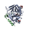
| Structure viewer | Molecule: MolmilJmol/JSmol |
|---|
- Downloads & links
-Download
| PDBx/mmCIF format | 3g2c.cif.gz | 129.3 KB | Display | PDBx/mmCIF format |
|---|---|---|---|---|
| PDB format | pdb3g2c.ent.gz | 98 KB | Display | PDB format |
| PDBx/mmJSON format | 3g2c.json.gz | Tree view | PDBx/mmJSON format | |
| Others |  Other downloads Other downloads |
-Validation report
| Arichive directory | https://data.pdbj.org/pub/pdb/validation_reports/g2/3g2cftp://data.pdbj.org/pub/pdb/validation_reports/g2/3g2c | HTTPS FTP |
|---|
-Related structure data
| Related structure data |  3fziSC  3g00C  3g0aC  3g0rC  3g1kC  3g2dC  3g38C  3g3cC  3g3yC  3g4tC  3g8vC  3g91C  3ga6C S: Starting model for refinement C: citing same article ( |
|---|---|
| Similar structure data |


-Links
 PDBj
PDBj


- Assembly
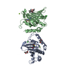
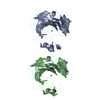
| Deposited unit | 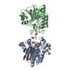
| ||||||||
|---|---|---|---|---|---|---|---|---|---|
| 1 |
| ||||||||
| Unit cell |
|
-Components
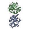
-Protein / DNA chain , 2 types, 3 molecules ABI
| #1: Protein | Mass: 31430.650 Da / Num. of mol.: 2 / Mutation: T2A Source method: isolated from a genetically manipulated source Source: (gene. exp.) Methanothermobacter thermautotrophicus (archaea)Strain: Delta H (DSM 1053) / Gene: mth0212, MTH212, MTH_212 / Plasmid: pET_B_001-mth212 (WT) / Production host:  #2: DNA chain | | Mass: 1190.830 Da / Num. of mol.: 1 / Source method: obtained synthetically |
|---|
-Non-polymers , 4 types, 180 molecules 


| #3: Chemical | ChemComp-GOL /  Mass: 92.094 Da / Num. of mol.: 7 / Source method: obtained synthetically / Formula: C3H8O3 Mass: 92.094 Da / Num. of mol.: 7 / Source method: obtained synthetically / Formula: C3H8O3#4: Chemical |  Mass: 24.305 Da / Num. of mol.: 2 / Source method: obtained synthetically / Formula: Mg Mass: 24.305 Da / Num. of mol.: 2 / Source method: obtained synthetically / Formula: Mg#5: Chemical |  Mass: 94.971 Da / Num. of mol.: 3 / Source method: obtained synthetically / Formula: PO4 Mass: 94.971 Da / Num. of mol.: 3 / Source method: obtained synthetically / Formula: PO4#6: Water | ChemComp-HOH / | Mass: 18.015 Da / Num. of mol.: 168 / Source method: isolated from a natural source / Formula: H2O |
|---|
-Details
| Sequence details | IN THIS ENTRY, THE FOLLOWING DNAS WERE PREPARED. THE ONE CONSISTS OF CGTA(UPS)TACG AND THE OTHER ...IN THIS ENTRY, THE FOLLOWING DNAS WERE PREPARED. THE ONE CONSISTS OF CGTA(UPS)TACG AND THE OTHER CGTATACG THAT WERE CONSTRUCTE |
|---|
-Experimental details
-Experiment
| Experiment | Method: X-RAY DIFFRACTION / Number of used crystals: 1 |
|---|
- Sample preparation
| Crystal | Density Matthews: 2.31 Å3/Da / Density % sol: 46.87 % | ||||||||||||||||||||||||||||||||
|---|---|---|---|---|---|---|---|---|---|---|---|---|---|---|---|---|---|---|---|---|---|---|---|---|---|---|---|---|---|---|---|---|---|
| Crystal grow | Temperature: 293 K / Method: vapor diffusion, sitting drop / pH: 7 Details: reservoir: 25% MPD, 100mM KHEPES pH 7.0; complex solution: 50mM KCl, 10mM KH2PO4/K2HPO4 pH 7.0, 1mM MgCl2, vapor diffusion, sitting drop, temperature 293K | ||||||||||||||||||||||||||||||||
| Components of the solutions |
|
-Data collection
| Diffraction | Mean temperature: 100 K |
|---|---|
| Diffraction source | Source: SYNCHROTRON / Site: EMBL/DESY, HAMBURG  / Beamline: X11 / Wavelength: 0.815 Å / Beamline: X11 / Wavelength: 0.815 Å |
| Detector | Type: MAR CCD 165 mm / Detector: CCD / Date: May 17, 2007 |
| Radiation | Protocol: SINGLE WAVELENGTH / Monochromatic (M) / Laue (L): M / Scattering type: x-ray |
| Radiation wavelength | Wavelength: 0.815 Å / Relative weight: 1 |
| Reflection | Resolution: 2.3→30 Å / Num. obs: 25521 / % possible obs: 99.8 % / Redundancy: 5.5 % / Biso Wilson estimate: 44.8 Å2 / Rsym value: 0.085 / Net I/σ(I): 17.1 |
| Reflection shell | Resolution: 2.3→2.38 Å / Redundancy: 4.5 % / Mean I/σ(I) obs: 4.1 / Rsym value: 0.347 / % possible all: 99.3 |
-Phasing
| Phasing | Method: molecular replacement |
|---|
- Processing
| Software |
| ||||||||||||||||||||||||||||||||||||||||||||||||||||||||||||||||||||||||||||||||||||||||||
|---|---|---|---|---|---|---|---|---|---|---|---|---|---|---|---|---|---|---|---|---|---|---|---|---|---|---|---|---|---|---|---|---|---|---|---|---|---|---|---|---|---|---|---|---|---|---|---|---|---|---|---|---|---|---|---|---|---|---|---|---|---|---|---|---|---|---|---|---|---|---|---|---|---|---|---|---|---|---|---|---|---|---|---|---|---|---|---|---|---|---|---|
| Refinement | Method to determine structure: MOLECULAR REPLACEMENT Starting model: PDB ENTRY 3FZI Resolution: 2.3→28.27 Å / Cor.coef. Fo:Fc: 0.943 / Cor.coef. Fo:Fc free: 0.906 / WRfactor Rfree: 0.274 / WRfactor Rwork: 0.209 / Occupancy max: 1 / Occupancy min: 0.5 / FOM work R set: 0.787 / SU B: 8.348 / SU ML: 0.206 / SU R Cruickshank DPI: 0.422 / SU Rfree: 0.268 / Cross valid method: THROUGHOUT / σ(F): 0 / ESU R: 0.422 / ESU R Free: 0.268 / Stereochemistry target values: MAXIMUM LIKELIHOOD Details: HYDROGENS HAVE BEEN ADDED IN THE RIDING POSITIONS; the coordinates of Thr 210 and Arg 211 of chain A are connected via a peptide bond which deviates significantly from both cis and trans ...Details: HYDROGENS HAVE BEEN ADDED IN THE RIDING POSITIONS; the coordinates of Thr 210 and Arg 211 of chain A are connected via a peptide bond which deviates significantly from both cis and trans conformation.; the coordinates representing the Arg 15 of chain A and Thr 210 of chain B have unexpected configuration of the chiral center. They should not be changed.
| ||||||||||||||||||||||||||||||||||||||||||||||||||||||||||||||||||||||||||||||||||||||||||
| Solvent computation | Ion probe radii: 0.8 Å / Shrinkage radii: 0.8 Å / VDW probe radii: 1.2 Å / Solvent model: MASK | ||||||||||||||||||||||||||||||||||||||||||||||||||||||||||||||||||||||||||||||||||||||||||
| Displacement parameters | Biso max: 83.95 Å2 / Biso mean: 38.834 Å2 / Biso min: 14.99 Å2
| ||||||||||||||||||||||||||||||||||||||||||||||||||||||||||||||||||||||||||||||||||||||||||
| Refinement step | Cycle: LAST / Resolution: 2.3→28.27 Å
| ||||||||||||||||||||||||||||||||||||||||||||||||||||||||||||||||||||||||||||||||||||||||||
| Refine LS restraints |
| ||||||||||||||||||||||||||||||||||||||||||||||||||||||||||||||||||||||||||||||||||||||||||
| LS refinement shell | Resolution: 2.299→2.359 Å / Total num. of bins used: 20
|
