 Movie
Movie Controller


+ Open data

- Basic information
| Entry | Database: PDB / ID: 3g0a | ||||||
|---|---|---|---|---|---|---|---|
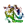
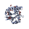
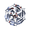
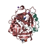
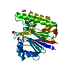
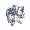
| Title | Mth0212 with two bound manganese ions | ||||||
 Components Components | Exodeoxyribonuclease | ||||||
 Keywords Keywords | HYDROLASE / coordination of two manganese ions / double-strand specific 3'-5' exonuclease / AP endonuclease / 2'-deoxyuridine endonuclease | ||||||
| Function / homology |  Function and homology information Function and homology informationexodeoxyribonuclease III / double-stranded DNA 3'-5' DNA exonuclease activity / phosphoric diester hydrolase activity / class I DNA-(apurinic or apyrimidinic site) endonuclease activity / DNA-(apurinic or apyrimidinic site) lyase / base-excision repair / DNA binding / metal ion binding Similarity search - Function | ||||||
| Biological species |   Methanothermobacter thermautotrophicus (archaea) Methanothermobacter thermautotrophicus (archaea) | ||||||
| Method |  X-RAY DIFFRACTION / MOLECULAR REPLACEMENT / molecular replacement / Resolution: 2.6 Å X-RAY DIFFRACTION / MOLECULAR REPLACEMENT / molecular replacement / Resolution: 2.6 Å | ||||||
 Authors Authors | Lakomek, K. / Dickmanns, A. / Ficner, R. | ||||||
 Citation Citation | Journal: J.Mol.Biol. / Year: 2010 Title: Crystal Structure Analysis of DNA Uridine Endonuclease Mth212 Bound to DNA Authors: Lakomek, K. / Dickmanns, A. / Ciirdaeva, E. / Schomacher, L. / Ficner, R. #1: Journal: Nucleic Acids Res. / Year: 2006 Title: The Methanothermobacter thermautotrophicus ExoIII homologue Mth212 is a DNA uridine endonuclease Authors: Georg, J. / Schomacher, L. / Chong, J.P.J. / Majernik, A.I. / Raabe, M. / Urlaub, H. / Muller, S. / Ciirdaeva, E. / Kramer, W. / Fritz, H.-J. | ||||||
| History |
|
- Structure visualization
| Structure viewer | Molecule: MolmilJmol/JSmol |
|---|
- Downloads & links
-Download
| PDBx/mmCIF format | 3g0a.cif.gz | 69.7 KB | Display | PDBx/mmCIF format |
|---|---|---|---|---|
| PDB format | pdb3g0a.ent.gz | 50.6 KB | Display | PDB format |
| PDBx/mmJSON format | 3g0a.json.gz | Tree view | PDBx/mmJSON format | |
| Others |  Other downloads Other downloads |
-Validation report
| Arichive directory | https://data.pdbj.org/pub/pdb/validation_reports/g0/3g0aftp://data.pdbj.org/pub/pdb/validation_reports/g0/3g0a | HTTPS FTP |
|---|
-Related structure data
| Related structure data |  3fziSC  3g00C  3g0rC  3g1kC  3g2cC  3g2dC  3g38C  3g3cC  3g3yC  3g4tC  3g8vC  3g91C  3ga6C S: Starting model for refinement C: citing same article ( |
|---|---|
| Similar structure data |


-Links
 PDBj
PDBj


- Assembly
| Deposited unit | 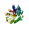
| ||||||||
|---|---|---|---|---|---|---|---|---|---|
| 1 |
| ||||||||
| Unit cell |
|
-Components
| #1: Protein | Mass: 31430.650 Da / Num. of mol.: 1 / Mutation: T2A Source method: isolated from a genetically manipulated source Details: lacking the gene "ung" Source: (gene. exp.) Methanothermobacter thermautotrophicus (archaea)Strain: Delta H (DSM 1053) / Gene: mth0212, MTH212, MTH_212 / Plasmid: pET_B_001-mth212 (WT) / Production host:  | ||||||
|---|---|---|---|---|---|---|---|
| #2: Chemical |   Mass: 54.938 Da / Num. of mol.: 2 / Source method: obtained synthetically / Formula: Mn Mass: 54.938 Da / Num. of mol.: 2 / Source method: obtained synthetically / Formula: Mn#3: Chemical | ChemComp-GOL / |   Mass: 92.094 Da / Num. of mol.: 1 / Source method: obtained synthetically / Formula: C3H8O3 Mass: 92.094 Da / Num. of mol.: 1 / Source method: obtained synthetically / Formula: C3H8O3#4: Chemical | ChemComp-PO4 / |   Mass: 94.971 Da / Num. of mol.: 1 / Source method: obtained synthetically / Formula: PO4 Mass: 94.971 Da / Num. of mol.: 1 / Source method: obtained synthetically / Formula: PO4#5: Water | ChemComp-HOH / |  Mass: 18.015 Da / Num. of mol.: 71 / Source method: isolated from a natural source / Formula: H2O Mass: 18.015 Da / Num. of mol.: 71 / Source method: isolated from a natural source / Formula: H2O |
-Experimental details
-Experiment
| Experiment | Method: X-RAY DIFFRACTION / Number of used crystals: 1 |
|---|
- Sample preparation
| Crystal | Density Matthews: 2.38 Å3/Da / Density % sol: 48.29 % |
|---|---|
| Crystal grow | Temperature: 293 K / Method: vapor diffusion, sitting drop / pH: 6.5 Details: reservoir solution: 10% (w/v) PEG 20000, 100mM MES-NaOH pH 6.5; protein solution: 600mM NaCl, 20mM HEPES-KOH pH 7.6, 2mM DTT; soaking in 6% (w/v) PEG 20000, 23% (v/v) glycerol, 60mM MES-NaOH ...Details: reservoir solution: 10% (w/v) PEG 20000, 100mM MES-NaOH pH 6.5; protein solution: 600mM NaCl, 20mM HEPES-KOH pH 7.6, 2mM DTT; soaking in 6% (w/v) PEG 20000, 23% (v/v) glycerol, 60mM MES-NaOH pH 6.5, 200mM MnCl2, VAPOR DIFFUSION, SITTING DROP, temperature 293K |
-Data collection
| Diffraction | Mean temperature: 130 K |
|---|---|
| Diffraction source | Source: ROTATING ANODE / Type: RIGAKU MICROMAX-007 / Wavelength: 1.5418 Å |
| Detector | Type: MAR scanner 345 mm plate / Detector: IMAGE PLATE / Date: Feb 28, 2007 |
| Radiation | Protocol: SINGLE WAVELENGTH / Monochromatic (M) / Laue (L): M / Scattering type: x-ray |
| Radiation wavelength | Wavelength: 1.5418 Å / Relative weight: 1 |
| Reflection | Resolution: 2.6→50 Å / Num. obs: 8864 / % possible obs: 96.6 % / Redundancy: 6.5 % / Rsym value: 0.095 / Net I/σ(I): 17 |
| Reflection shell | Resolution: 2.6→2.69 Å / Redundancy: 2.4 % / Rsym value: 0.351 |
-Phasing
| Phasing | Method: molecular replacement |
|---|
- Processing
| Software |
| ||||||||||||||||||||||||||||||||||||||||||||||||||||||||||||||||||||||||||||||||||||||||||
|---|---|---|---|---|---|---|---|---|---|---|---|---|---|---|---|---|---|---|---|---|---|---|---|---|---|---|---|---|---|---|---|---|---|---|---|---|---|---|---|---|---|---|---|---|---|---|---|---|---|---|---|---|---|---|---|---|---|---|---|---|---|---|---|---|---|---|---|---|---|---|---|---|---|---|---|---|---|---|---|---|---|---|---|---|---|---|---|---|---|---|---|
| Refinement | Method to determine structure: MOLECULAR REPLACEMENT Starting model: PDB ENTRY 3FZI Resolution: 2.6→48.83 Å / Cor.coef. Fo:Fc: 0.938 / Cor.coef. Fo:Fc free: 0.896 / Occupancy max: 1 / Occupancy min: 0.5 / SU B: 11.573 / SU ML: 0.245 / Cross valid method: THROUGHOUT / σ(F): 0 / ESU R Free: 0.345 / Stereochemistry target values: MAXIMUM LIKELIHOOD / Details: HYDROGENS HAVE BEEN ADDED IN THE RIDING POSITIONS
| ||||||||||||||||||||||||||||||||||||||||||||||||||||||||||||||||||||||||||||||||||||||||||
| Solvent computation | Ion probe radii: 0.8 Å / Shrinkage radii: 0.8 Å / VDW probe radii: 1.2 Å / Solvent model: MASK | ||||||||||||||||||||||||||||||||||||||||||||||||||||||||||||||||||||||||||||||||||||||||||
| Displacement parameters | Biso max: 75.26 Å2 / Biso mean: 32.202 Å2 / Biso min: 9.09 Å2
| ||||||||||||||||||||||||||||||||||||||||||||||||||||||||||||||||||||||||||||||||||||||||||
| Refinement step | Cycle: LAST / Resolution: 2.6→48.83 Å
| ||||||||||||||||||||||||||||||||||||||||||||||||||||||||||||||||||||||||||||||||||||||||||
| Refine LS restraints |
| ||||||||||||||||||||||||||||||||||||||||||||||||||||||||||||||||||||||||||||||||||||||||||
| LS refinement shell | Resolution: 2.603→2.67 Å / Total num. of bins used: 20
|
