 Movie
Movie Controller


+ Open data

- Basic information
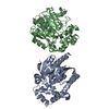
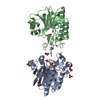
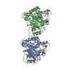
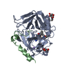
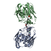
| Entry | Database: PDB / ID: 3g0r | ||||||
|---|---|---|---|---|---|---|---|
| Title | Complex of Mth0212 and an 8bp dsDNA with distorted ends | ||||||
 Components Components |
| ||||||
 Keywords Keywords | HYDROLASE/DNA / protein-DNA complex / double-strand specific 3'-5' exonuclease / AP endonuclease / 2'-deoxyuridine endonuclease / HYDROLASE-DNA COMPLEX | ||||||
| Function / homology |  Function and homology information Function and homology informationexodeoxyribonuclease III / double-stranded DNA 3'-5' DNA exonuclease activity / phosphoric diester hydrolase activity / class I DNA-(apurinic or apyrimidinic site) endonuclease activity / DNA-(apurinic or apyrimidinic site) lyase / base-excision repair / DNA binding / metal ion binding Similarity search - Function | ||||||
| Biological species |   Methanothermobacter thermautotrophicus (archaea) Methanothermobacter thermautotrophicus (archaea) | ||||||
| Method |  X-RAY DIFFRACTION / SYNCHROTRON / MOLECULAR REPLACEMENT / molecular replacement / Resolution: 2.4 Å X-RAY DIFFRACTION / SYNCHROTRON / MOLECULAR REPLACEMENT / molecular replacement / Resolution: 2.4 Å | ||||||
 Authors Authors | Lakomek, K. / Dickmanns, A. / Ficner, R. | ||||||
 Citation Citation | Journal: J.Mol.Biol. / Year: 2010 Title: Crystal Structure Analysis of DNA Uridine Endonuclease Mth212 Bound to DNA Authors: Lakomek, K. / Dickmanns, A. / Ciirdaeva, E. / Schomacher, L. / Ficner, R. #1: Journal: Nucleic Acids Res. / Year: 2006 Title: The Methanothermobacter thermautotrophicus ExoIII homologue Mth212 is a DNA uridine endonuclease Authors: Georg, J. / Schomacher, L. / Chong, J.P.J. / Majernik, A.I. / Raabe, M. / Urlaub, H. / Muller, S. / Ciirdaeva, E. / Kramer, W. / Fritz, H.-J. | ||||||
| History |
|
- Structure visualization
| Structure viewer | Molecule: MolmilJmol/JSmol |
|---|
- Downloads & links
-Download
| PDBx/mmCIF format | 3g0r.cif.gz | 145.4 KB | Display | PDBx/mmCIF format |
|---|---|---|---|---|
| PDB format | pdb3g0r.ent.gz | 109.3 KB | Display | PDB format |
| PDBx/mmJSON format | 3g0r.json.gz | Tree view | PDBx/mmJSON format | |
| Others |  Other downloads Other downloads |
-Validation report
| Arichive directory | https://data.pdbj.org/pub/pdb/validation_reports/g0/3g0rftp://data.pdbj.org/pub/pdb/validation_reports/g0/3g0r | HTTPS FTP |
|---|
-Related structure data
| Related structure data |  3fziSC  3g00C  3g0aC  3g1kC  3g2cC  3g2dC  3g38C  3g3cC  3g3yC  3g4tC  3g8vC  3g91C  3ga6C S: Starting model for refinement C: citing same article ( |
|---|---|
| Similar structure data |


-Links
 PDBj
PDBj


- Assembly
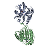
| Deposited unit | 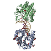
| ||||||||
|---|---|---|---|---|---|---|---|---|---|
| 1 |
| ||||||||
| Unit cell |
|
-Components
-Protein , 1 types, 2 molecules AB
| #1: Protein | Mass: 31429.666 Da / Num. of mol.: 2 / Mutation: T2A, D151N Source method: isolated from a genetically manipulated source Source: (gene. exp.) Methanothermobacter thermautotrophicus (archaea)Strain: Delta H (DSM 1053) / Gene: mth0212, MTH212, MTH_212 / Plasmid: pET_B_001-mth212 (D151N) / Production host:  |
|---|
-DNA chain , 2 types, 2 molecules GK
| #2: DNA chain | Mass: 4033.609 Da / Num. of mol.: 1 / Source method: obtained synthetically |
|---|---|
| #3: DNA chain | Mass: 3296.135 Da / Num. of mol.: 1 / Source method: obtained synthetically |
-Non-polymers , 4 types, 322 molecules 


| #4: Chemical | ChemComp-GOL /  Mass: 92.094 Da / Num. of mol.: 12 / Source method: obtained synthetically / Formula: C3H8O3 Mass: 92.094 Da / Num. of mol.: 12 / Source method: obtained synthetically / Formula: C3H8O3#5: Chemical |  Mass: 194.226 Da / Num. of mol.: 3 / Source method: obtained synthetically / Formula: C8H18O5 / Comment: precipitant*YM Mass: 194.226 Da / Num. of mol.: 3 / Source method: obtained synthetically / Formula: C8H18O5 / Comment: precipitant*YM#6: Chemical |  Mass: 22.990 Da / Num. of mol.: 2 / Source method: obtained synthetically / Formula: Na Mass: 22.990 Da / Num. of mol.: 2 / Source method: obtained synthetically / Formula: Na#7: Water | ChemComp-HOH / | Mass: 18.015 Da / Num. of mol.: 305 / Source method: isolated from a natural source / Formula: H2O |
|---|
-Details
| Compound details | THE 2ND DC IN CHAIN K STANDS FOR THE MODIFIED NUCLEOTIDE 5- BROMO-2'DC. SINCE THERE WAS NO HINT FOR ...THE 2ND DC IN CHAIN K STANDS FOR THE MODIFIED NUCLEOTIDE |
|---|
-Experimental details
-Experiment
| Experiment | Method: X-RAY DIFFRACTION / Number of used crystals: 1 |
|---|
- Sample preparation
| Crystal | Density Matthews: 2.7 Å3/Da / Density % sol: 54.48 % | ||||||||||||||||||||||||||||||||||||||||||||||||||||
|---|---|---|---|---|---|---|---|---|---|---|---|---|---|---|---|---|---|---|---|---|---|---|---|---|---|---|---|---|---|---|---|---|---|---|---|---|---|---|---|---|---|---|---|---|---|---|---|---|---|---|---|---|---|
| Crystal grow | Temperature: 293 K / Method: vapor diffusion, sitting drop / pH: 5.6 Details: reservoir: 189mM KCl, 9.5mM MgCl2, 4.7% (w/v) PEG 8000, 47mM MES/NaOH pH 5.6, 2.1% 1,4-butanediol, 2mM DTT; protein solution: 234mM NaCl, 7mM KHEPES pH 7.6, 1.83mM MgCl2, 2.7mM DTT, VAPOR ...Details: reservoir: 189mM KCl, 9.5mM MgCl2, 4.7% (w/v) PEG 8000, 47mM MES/NaOH pH 5.6, 2.1% 1,4-butanediol, 2mM DTT; protein solution: 234mM NaCl, 7mM KHEPES pH 7.6, 1.83mM MgCl2, 2.7mM DTT, VAPOR DIFFUSION, SITTING DROP, temperature 293K | ||||||||||||||||||||||||||||||||||||||||||||||||||||
| Components of the solutions |
|
-Data collection
| Diffraction | Mean temperature: 100 K |
|---|---|
| Diffraction source | Source: SYNCHROTRON / Site: EMBL/DESY, HAMBURG  / Beamline: X12 / Wavelength: 0.97623 Å / Beamline: X12 / Wavelength: 0.97623 Å |
| Detector | Type: MARMOSAIC 225 mm CCD / Detector: CCD / Date: Feb 5, 2007 |
| Radiation | Protocol: SINGLE WAVELENGTH / Monochromatic (M) / Laue (L): M / Scattering type: x-ray |
| Radiation wavelength | Wavelength: 0.97623 Å / Relative weight: 1 |
| Reflection | Resolution: 2.4→50 Å / Num. obs: 27308 / % possible obs: 92.8 % / Redundancy: 4.7 % / Rsym value: 0.061 / Net I/σ(I): 22.7 |
| Reflection shell | Resolution: 2.4→2.49 Å / Redundancy: 3.2 % / Mean I/σ(I) obs: 5.2 / Rsym value: 0.191 / % possible all: 72.3 |
-Phasing
| Phasing | Method: molecular replacement |
|---|
- Processing
| Software |
| ||||||||||||||||||||||||||||||||||||||||||||||||||||||||||||||||||||||||||||||||||||||||||||||||||||||||||||||||||||||||||||||||||||||||||||||||||||||||||||||||||||||||||
|---|---|---|---|---|---|---|---|---|---|---|---|---|---|---|---|---|---|---|---|---|---|---|---|---|---|---|---|---|---|---|---|---|---|---|---|---|---|---|---|---|---|---|---|---|---|---|---|---|---|---|---|---|---|---|---|---|---|---|---|---|---|---|---|---|---|---|---|---|---|---|---|---|---|---|---|---|---|---|---|---|---|---|---|---|---|---|---|---|---|---|---|---|---|---|---|---|---|---|---|---|---|---|---|---|---|---|---|---|---|---|---|---|---|---|---|---|---|---|---|---|---|---|---|---|---|---|---|---|---|---|---|---|---|---|---|---|---|---|---|---|---|---|---|---|---|---|---|---|---|---|---|---|---|---|---|---|---|---|---|---|---|---|---|---|---|---|---|---|---|---|---|
| Refinement | Method to determine structure: MOLECULAR REPLACEMENT Starting model: PDB ENTRY 3FZI Resolution: 2.4→44.64 Å / Cor.coef. Fo:Fc: 0.943 / Cor.coef. Fo:Fc free: 0.906 / WRfactor Rfree: 0.226 / WRfactor Rwork: 0.171 / Occupancy max: 1 / Occupancy min: 0.5 / FOM work R set: 0.824 / SU B: 7.338 / SU ML: 0.174 / SU R Cruickshank DPI: 0.427 / SU Rfree: 0.271 / Cross valid method: THROUGHOUT / σ(F): 0 / ESU R: 0.427 / ESU R Free: 0.27 / Stereochemistry target values: MAXIMUM LIKELIHOOD / Details: HYDROGENS HAVE BEEN ADDED IN THE RIDING POSITIONS
| ||||||||||||||||||||||||||||||||||||||||||||||||||||||||||||||||||||||||||||||||||||||||||||||||||||||||||||||||||||||||||||||||||||||||||||||||||||||||||||||||||||||||||
| Solvent computation | Ion probe radii: 0.8 Å / Shrinkage radii: 0.8 Å / VDW probe radii: 1.2 Å / Solvent model: MASK | ||||||||||||||||||||||||||||||||||||||||||||||||||||||||||||||||||||||||||||||||||||||||||||||||||||||||||||||||||||||||||||||||||||||||||||||||||||||||||||||||||||||||||
| Displacement parameters | Biso max: 71.56 Å2 / Biso mean: 27.829 Å2 / Biso min: 4.11 Å2
| ||||||||||||||||||||||||||||||||||||||||||||||||||||||||||||||||||||||||||||||||||||||||||||||||||||||||||||||||||||||||||||||||||||||||||||||||||||||||||||||||||||||||||
| Refinement step | Cycle: LAST / Resolution: 2.4→44.64 Å
| ||||||||||||||||||||||||||||||||||||||||||||||||||||||||||||||||||||||||||||||||||||||||||||||||||||||||||||||||||||||||||||||||||||||||||||||||||||||||||||||||||||||||||
| Refine LS restraints |
| ||||||||||||||||||||||||||||||||||||||||||||||||||||||||||||||||||||||||||||||||||||||||||||||||||||||||||||||||||||||||||||||||||||||||||||||||||||||||||||||||||||||||||
| LS refinement shell | Resolution: 2.399→2.461 Å / Total num. of bins used: 20
|
