[English] 日本語
Yorodumi
- PDB-2j0e: Three dimensional structure and catalytic mechanism of 6- phospho... -

+
Open data


ID or keywords:

Loading...

-
Basic information

Entry
Database: PDB / ID: 2j0e
TitleThree dimensional structure and catalytic mechanism of 6- phosphogluconolactonase from Trypanosoma brucei
Components6-PHOSPHOGLUCONOLACTONASE
KeywordsHYDROLASE / CATALYTIC MECHANISM / PENTOSE PHOSPHATE PATHWAY / 6-PHOSPHOGLUCONOLACTONASE / TRYPANOSOMA BRUCEI / ZINC BINDING SITE
Function / homology
Function and homology information


6-phosphogluconolactonase / 6-phosphogluconolactonase activity / glycosome / pentose-phosphate shunt / carbohydrate metabolic process / metal ion binding / cytoplasm
Similarity search - Function
6-Phosphogluconolactonase / 6-phosphogluconolactonase, DevB-type / Glucosamine/galactosamine-6-phosphate isomerase / Glucosamine-6-phosphate isomerases/6-phosphogluconolactonase / Rossmann fold - #1360 / NagB/RpiA transferase-like / Rossmann fold / 3-Layer(aba) Sandwich / Alpha Beta
Similarity search - Domain/homology
: / : / 6-phosphogluconolactonase
Similarity search - Component
Biological speciesTRYPANOSOMA BRUCEI (eukaryote)
MethodX-RAY DIFFRACTION / SYNCHROTRON / SAD / Resolution: 2.1 Å
AuthorsDelarue, M. / Duclert-Savatier, N. / Miclet, E. / Haouz, A. / Giganti, D. / Ouazzani, J. / Lopez, P. / Nilges, M. / Stoven, V.
CitationJournal: J.Mol.Biol. / Year: 2007
Title: Three Dimensional Structure and Implications for the Catalytic Mechanism of 6-Phosphogluconolactonase from Trypanosoma Brucei.
Authors: Delarue, M. / Duclert-Savatier, N. / Miclet, E. / Haouz, A. / Giganti, D. / Ouazzani, J. / Lopez, P. / Nilges, M. / Stoven, V.
History
DepositionAug 2, 2006Deposition site: PDBE / Processing site: PDBE
Revision 1.0Jan 3, 2007Provider: repository / Type: Initial release
Revision 1.1May 8, 2011Group: Version format compliance
Revision 1.2Jul 13, 2011Group: Version format compliance
Revision 1.3May 8, 2024Group: Data collection / Database references ...Data collection / Database references / Derived calculations / Other
Category: chem_comp_atom / chem_comp_bond ...chem_comp_atom / chem_comp_bond / database_2 / pdbx_database_status / pdbx_struct_conn_angle / struct_conn
Item: _database_2.pdbx_DOI / _database_2.pdbx_database_accession ..._database_2.pdbx_DOI / _database_2.pdbx_database_accession / _pdbx_database_status.status_code_sf / _pdbx_struct_conn_angle.ptnr1_auth_comp_id / _pdbx_struct_conn_angle.ptnr1_auth_seq_id / _pdbx_struct_conn_angle.ptnr1_label_asym_id / _pdbx_struct_conn_angle.ptnr1_label_atom_id / _pdbx_struct_conn_angle.ptnr1_label_comp_id / _pdbx_struct_conn_angle.ptnr1_label_seq_id / _pdbx_struct_conn_angle.ptnr2_auth_comp_id / _pdbx_struct_conn_angle.ptnr2_auth_seq_id / _pdbx_struct_conn_angle.ptnr2_label_asym_id / _pdbx_struct_conn_angle.ptnr2_label_atom_id / _pdbx_struct_conn_angle.ptnr2_label_comp_id / _pdbx_struct_conn_angle.ptnr3_auth_comp_id / _pdbx_struct_conn_angle.ptnr3_auth_seq_id / _pdbx_struct_conn_angle.ptnr3_label_asym_id / _pdbx_struct_conn_angle.ptnr3_label_atom_id / _pdbx_struct_conn_angle.ptnr3_label_comp_id / _pdbx_struct_conn_angle.ptnr3_label_seq_id / _pdbx_struct_conn_angle.value / _struct_conn.pdbx_dist_value / _struct_conn.ptnr1_auth_comp_id / _struct_conn.ptnr1_auth_seq_id / _struct_conn.ptnr1_label_asym_id / _struct_conn.ptnr1_label_atom_id / _struct_conn.ptnr1_label_comp_id / _struct_conn.ptnr1_label_seq_id / _struct_conn.ptnr2_auth_comp_id / _struct_conn.ptnr2_auth_seq_id / _struct_conn.ptnr2_label_asym_id / _struct_conn.ptnr2_label_atom_id / _struct_conn.ptnr2_label_comp_id / _struct_conn.ptnr2_label_seq_id

-
Structure visualization

Structure viewerMolecule:
MolmilJmol/JSmol

Downloads & links

-
Assembly

Deposited unit
A: 6-PHOSPHOGLUCONOLACTONASE
B: 6-PHOSPHOGLUCONOLACTONASE
hetero molecules


Theoretical massNumber of molelcules
Total (without water)58,3329
Polymers57,3602
Non-polymers9727
Water8,305461
1
A: 6-PHOSPHOGLUCONOLACTONASE
hetero molecules


Theoretical massNumber of molelcules
Total (without water)29,1464
Polymers28,6801
Non-polymers4673
Water181
TypeNameSymmetry operationNumber
identity operation1_555x,y,z1
MethodPQS
2
B: 6-PHOSPHOGLUCONOLACTONASE
hetero molecules


Theoretical massNumber of molelcules
Total (without water)29,1865
Polymers28,6801
Non-polymers5064
Water181
TypeNameSymmetry operationNumber
identity operation1_555x,y,z1
MethodPQS
Unit cell
Length a, b, c (Å)70.310, 80.850, 90.310
Angle α, β, γ (deg.)90.00, 90.00, 90.00
Int Tables number19
Space group name H-MP212121
Noncrystallographic symmetry (NCS)NCS oper: (Code: given
Matrix: (0.9994, -0.0079, -0.0347), (0.0108, 0.9964, 0.0842), (0.0339, -0.0845, 0.9958)
Vector: -0.3937, 14.3518, -46.0985)

-
Components

#1: Protein 6-PHOSPHOGLUCONOLACTONASE


Mass: 28679.891 Da / Num. of mol.: 2
Source method: isolated from a genetically manipulated source
Details: ONE BOUND ZINC ATOM PER MONOMER. / Source: (gene. exp.) TRYPANOSOMA BRUCEI (eukaryote) / Plasmid: PET21 / Production host: ESCHERICHIA COLI (E. coli) / Strain (production host): BL21 / References: UniProt: Q9GRG6, 6-phosphogluconolactonase
#2: Chemical ChemComp-ZN / ZINC ION


Mass: 65.409 Da / Num. of mol.: 2 / Source method: obtained synthetically / Formula: Zn
#3: Chemical
ChemComp-HG / MERCURY (II) ION


Mass: 200.590 Da / Num. of mol.: 4 / Source method: obtained synthetically / Formula: Hg
#4: Chemical ChemComp-K / POTASSIUM ION


Mass: 39.098 Da / Num. of mol.: 1 / Source method: obtained synthetically / Formula: K
#5: Water ChemComp-HOH / water


Mass: 18.015 Da / Num. of mol.: 461 / Source method: isolated from a natural source / Formula: H2O

-
Experimental details

-
Experiment

ExperimentMethod: X-RAY DIFFRACTION / Number of used crystals: 1

-
Sample preparation

CrystalDensity Matthews: 2.32 Å3/Da / Density % sol: 46 %
Description: ANOTHER DATASET WITH SELENOMETHIONYLATED PROTEIN WAS COLLECTED AT 2.8 ANGSTROM AND WAS USED TO CHECK THE POSITION OF METHIONINES.
Crystal growpH: 7.5
Details: 30% PEG MME, 50MM NA ACETATE, HEPES PH 7.5 100 MM. PROTEIN 10MG/ML

-
Data collection

DiffractionMean temperature: 110 K
Diffraction sourceSource: SYNCHROTRON / Site: ESRF / Beamline: BM30A / Wavelength: 1.008014
DetectorType: MARRESEARCH / Detector: CCD / Date: Apr 15, 2004
RadiationProtocol: SINGLE WAVELENGTH / Monochromatic (M) / Laue (L): M / Scattering type: x-ray
Radiation wavelengthWavelength: 1.008014 Å / Relative weight: 1
ReflectionResolution: 2.1→100 Å / Num. obs: 57076 / % possible obs: 98.9 % / Observed criterion σ(I): 0 / Redundancy: 3.84 % / Biso Wilson estimate: 16.9 Å2 / Rmerge(I) obs: 0.06 / Net I/σ(I): 15.92
Reflection shellResolution: 2.1→2.2 Å / Redundancy: 3.5 % / Rmerge(I) obs: 0.24 / Mean I/σ(I) obs: 5.86 / % possible all: 99.3

-
Processing

Software
NameVersionClassification
CNS1.1refinement
MOSFLMdata reduction
SCALEPACKdata scaling
SOLVEphasing
Refinement
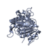
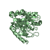
Method to determine structure: SAD / Resolution: 2.1→18 Å / Rfactor Rfree error: 0.006 / Data cutoff high absF: 10000000 / Isotropic thermal model: RESTRAINED / Cross valid method: THROUGHOUT / σ(F): 0 / Stereochemistry target values: MAXIMUM LIKELIHOOD
Details: THE STRUCTURE WAS REFINED ON THE CRYSTAL SOAKED WITH A MERCURY DERIVATIVE (2 HG ATOMS PER MONOMER)
RfactorNum. reflection% reflectionSelection details
Rfree0.246 1539 5.1 %RANDOM
Rwork0.206 ---
obs0.206 30438 99.3 %-
Solvent computationSolvent model: FLAT MODEL / Bsol: 65.7024 Å2 / ksol: 0.334866 e/Å3
Displacement parametersBiso mean: 28.4 Å2
Baniso -1Baniso -2Baniso -3
1--0.997 Å20 Å20 Å2
2--2.535 Å20 Å2
3----1.538 Å2
Refine analyze
FreeObs
Luzzati coordinate error0.3 Å0.23 Å
Luzzati d res low-5 Å
Luzzati sigma a0.25 Å0.18 Å
Refinement stepCycle: LAST / Resolution: 2.1→18 Å
ProteinNucleic acidLigandSolventTotal
Num. atoms3940 0 7 461 4408
Refine LS restraints
Refine-IDTypeDev idealDev ideal target
X-RAY DIFFRACTIONc_bond_d0.006
X-RAY DIFFRACTIONc_bond_d_na
X-RAY DIFFRACTIONc_bond_d_prot
X-RAY DIFFRACTIONc_angle_d
X-RAY DIFFRACTIONc_angle_d_na
X-RAY DIFFRACTIONc_angle_d_prot
X-RAY DIFFRACTIONc_angle_deg1.3
X-RAY DIFFRACTIONc_angle_deg_na
X-RAY DIFFRACTIONc_angle_deg_prot
X-RAY DIFFRACTIONc_dihedral_angle_d23
X-RAY DIFFRACTIONc_dihedral_angle_d_na
X-RAY DIFFRACTIONc_dihedral_angle_d_prot
X-RAY DIFFRACTIONc_improper_angle_d0.85
X-RAY DIFFRACTIONc_improper_angle_d_na
X-RAY DIFFRACTIONc_improper_angle_d_prot
X-RAY DIFFRACTIONc_mcbond_it1.371.5
X-RAY DIFFRACTIONc_mcangle_it2.162
X-RAY DIFFRACTIONc_scbond_it2.192
X-RAY DIFFRACTIONc_scangle_it3.182.5
LS refinement shellResolution: 2.1→2.23 Å / Rfactor Rfree error: 0.018 / Total num. of bins used: 6
RfactorNum. reflection% reflection
Rfree0.282 240 5 %
Rwork0.225 4603 -
obs--96.1 %
Xplor file
Refine-IDSerial noParam fileTopol file
X-RAY DIFFRACTION1PROTEIN_REP.PARAMPROTEIN.TOP
X-RAY DIFFRACTION2WATER_REP.PARAMWATER.TOP
X-RAY DIFFRACTION3ION.PARAMION.TOP
X-RAY DIFFRACTION4CIS_PEPTIDE.PARAM

+
About Yorodumi

-
News

-
Feb 9, 2022. New format data for meta-information of EMDB entries

New format data for meta-information of EMDB entries

  • Version 3 of the EMDB header file is now the official format.
  • The previous official version 1.9 will be removed from the archive.

Related info.:EMDB header

External links:wwPDB to switch to version 3 of the EMDB data model

-
Aug 12, 2020. Covid-19 info

Covid-19 info

URL: https://pdbj.org/emnavi/covid19.php

New page: Covid-19 featured information page in EM Navigator.

Related info.:Covid-19 info / Mar 5, 2020. Novel coronavirus structure data

+
Mar 5, 2020. Novel coronavirus structure data

Novel coronavirus structure data

Related info.:Yorodumi Speices / Aug 12, 2020. Covid-19 info

External links:COVID-19 featured content - PDBj / Molecule of the Month (242):Coronavirus Proteases

+
Jan 31, 2019. EMDB accession codes are about to change! (news from PDBe EMDB page)

EMDB accession codes are about to change! (news from PDBe EMDB page)

  • The allocation of 4 digits for EMDB accession codes will soon come to an end. Whilst these codes will remain in use, new EMDB accession codes will include an additional digit and will expand incrementally as the available range of codes is exhausted. The current 4-digit format prefixed with “EMD-” (i.e. EMD-XXXX) will advance to a 5-digit format (i.e. EMD-XXXXX), and so on. It is currently estimated that the 4-digit codes will be depleted around Spring 2019, at which point the 5-digit format will come into force.
  • The EM Navigator/Yorodumi systems omit the EMD- prefix.

Related info.:Q: What is EMD? / ID/Accession-code notation in Yorodumi/EM Navigator

External links:EMDB Accession Codes are Changing Soon! / Contact to PDBj

+
Jul 12, 2017. Major update of PDB

Major update of PDB

  • wwPDB released updated PDB data conforming to the new PDBx/mmCIF dictionary.
  • This is a major update changing the version number from 4 to 5, and with Remediation, in which all the entries are updated.
  • In this update, many items about electron microscopy experimental information are reorganized (e.g. em_software).
  • Now, EM Navigator and Yorodumi are based on the updated data.

External links:wwPDB Remediation / Enriched Model Files Conforming to OneDep Data Standards Now Available in the PDB FTP Archive

-
Yorodumi

Thousand views of thousand structures

  • Yorodumi is a browser for structure data from EMDB, PDB, SASBDB, etc.
  • This page is also the successor to EM Navigator detail page, and also detail information page/front-end page for Omokage search.
  • The word "yorodu" (or yorozu) is an old Japanese word meaning "ten thousand". "mi" (miru) is to see.

Related info.:EMDB / PDB / SASBDB / Comparison of 3 databanks / Yorodumi Search / Aug 31, 2016. New EM Navigator & Yorodumi / Yorodumi Papers / Jmol/JSmol / Function and homology information / Changes in new EM Navigator and Yorodumi

Read more