 Movie
Movie Controller


+ Open data

- Basic information
| Entry | Database: PDB / ID: 2voa | ||||||
|---|---|---|---|---|---|---|---|
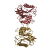
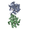
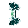
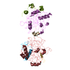
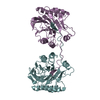
| Title | Structure of an AP Endonuclease from Archaeoglobus fulgidus | ||||||
 Components Components |
| ||||||
 Keywords Keywords | LYASE / EXOIII / AP ENDONUCLEASE / ARCHAEOGLOBUS FULGIDUS | ||||||
| Function / homology |  Function and homology information Function and homology informationdouble-stranded DNA 3'-5' DNA exonuclease activity / endonuclease activity / DNA repair / DNA binding / metal ion binding Similarity search - Function | ||||||
| Biological species |   ARCHAEOGLOBUS FULGIDUS (archaea) ARCHAEOGLOBUS FULGIDUS (archaea)synthetic construct (others) | ||||||
| Method |  X-RAY DIFFRACTION / SYNCHROTRON / MOLECULAR REPLACEMENT / Resolution: 1.7 Å X-RAY DIFFRACTION / SYNCHROTRON / MOLECULAR REPLACEMENT / Resolution: 1.7 Å | ||||||
 Authors Authors | Kuettner, E.B. / Schmiedel, R. / Greiner-Stoffele, T. / Strater, N. | ||||||
 Citation Citation | Journal: DNA Repair / Year: 2009 Title: Structure and Function of the Abasic Site Specificity Pocket of an Ap Endonuclease from Archaeoglobus Fulgidus. Authors: Schmiedel, R. / Kuettner, E.B. / Keim, A. / Strater, N. / Greiner-Stoffele, T. #1: Journal: Acta Crystallogr.,Sect.F / Year: 2006 Title: Crystallization and Preliminary X-Ray Characterization of Two Thermostable DNA Nucleases Authors: Kuettner, E.B. / Pfeifer, S. / Keim, A. / Greiner-Stoffele, T. / Strater, N. | ||||||
| History |
| ||||||
| Remark 650 | HELIX DETERMINATION METHOD: AUTHOR PROVIDED. | ||||||
| Remark 700 | SHEET DETERMINATION METHOD: AUTHOR PROVIDED. |
- Structure visualization
| Structure viewer | Molecule: MolmilJmol/JSmol |
|---|
- Downloads & links
-Download
| PDBx/mmCIF format | 2voa.cif.gz | 133.6 KB | Display | PDBx/mmCIF format |
|---|---|---|---|---|
| PDB format | pdb2voa.ent.gz | 100.8 KB | Display | PDB format |
| PDBx/mmJSON format | 2voa.json.gz | Tree view | PDBx/mmJSON format | |
| Others |  Other downloads Other downloads |
-Validation report
| Arichive directory | https://data.pdbj.org/pub/pdb/validation_reports/vo/2voaftp://data.pdbj.org/pub/pdb/validation_reports/vo/2voa | HTTPS FTP |
|---|
-Related structure data
| Related structure data |  1akoS S: Starting model for refinement |
|---|---|
| Similar structure data |


-Links
 PDBj
PDBj

- Assembly
| Deposited unit | 
| ||||||||
|---|---|---|---|---|---|---|---|---|---|
| 1 |
| ||||||||
| Unit cell |
|
-Components
| #1: Protein | Mass: 29810.215 Da / Num. of mol.: 2 / Mutation: YES Source method: isolated from a genetically manipulated source Source: (gene. exp.) ARCHAEOGLOBUS FULGIDUS (archaea) / Description: DSM4304 / Plasmid: PET28 / Production host:  References: UniProt: O29675, DNA-(apurinic or apyrimidinic site) lyase #2: DNA chain | | Mass: 3086.017 Da / Num. of mol.: 1 / Source method: obtained synthetically / Source: (synth.) synthetic construct (others) #3: DNA chain | | Mass: 3005.969 Da / Num. of mol.: 1 / Source method: obtained synthetically / Source: (synth.) synthetic construct (others) #4: Water | ChemComp-HOH / |  Mass: 18.015 Da / Num. of mol.: 220 / Source method: isolated from a natural source / Formula: H2O Mass: 18.015 Da / Num. of mol.: 220 / Source method: isolated from a natural source / Formula: H2OCompound details | ENGINEERED | Sequence details | V217G | |
|---|
-Experimental details
-Experiment
| Experiment | Method: X-RAY DIFFRACTION / Number of used crystals: 1 |
|---|
- Sample preparation
| Crystal | Density Matthews: 2.2 Å3/Da / Density % sol: 43.67 % / Description: NONE |
|---|---|
| Crystal grow | Temperature: 292 K / Method: vapor diffusion, hanging drop / pH: 7.8 Details: HANGING-DROP METHOD AT 292 K WITH 1 MICROLITER OF RESERVOIR AND PROTEIN SOLUTION EACH. TRIS-HCL PH 8.5. PROTEIN: 17 MG/ML AF_EXO, 0.53 MM DS-DNA, 9 MM MES-NAOH PH 6, 0.28 M NACL. |
-Data collection
| Diffraction | Mean temperature: 100 K |
|---|---|
| Diffraction source | Source: SYNCHROTRON / Site: BESSY  / Beamline: 14.2 / Wavelength: 0.9537 / Beamline: 14.2 / Wavelength: 0.9537 |
| Detector | Type: MARRESEARCH / Detector: CCD / Date: Feb 10, 2005 / Details: MIRRORS |
| Radiation | Monochromator: SI-111 CRYSTAL / Protocol: SINGLE WAVELENGTH / Monochromatic (M) / Laue (L): M / Scattering type: x-ray |
| Radiation wavelength | Wavelength: 0.9537 Å / Relative weight: 1 |
| Reflection | Resolution: 1.7→30 Å / Num. obs: 64871 / % possible obs: 100 % / Observed criterion σ(I): -3 / Redundancy: 7.8 % / Biso Wilson estimate: 29.5 Å2 / Rmerge(I) obs: 0.07 / Net I/σ(I): 24.5 |
| Reflection shell | Resolution: 1.7→1.74 Å / Redundancy: 5.4 % / Rmerge(I) obs: 0.44 / Mean I/σ(I) obs: 2.3 / % possible all: 99.9 |
- Processing
| Software |
| ||||||||||||||||||||||||||||||||||||||||||||||||||||||||||||||||||||||||||||||||||||||||||||||||||||||||||||||||||||||||||||||||||||||||||||||||||||||||||||||||||||||||||||||||||||||
|---|---|---|---|---|---|---|---|---|---|---|---|---|---|---|---|---|---|---|---|---|---|---|---|---|---|---|---|---|---|---|---|---|---|---|---|---|---|---|---|---|---|---|---|---|---|---|---|---|---|---|---|---|---|---|---|---|---|---|---|---|---|---|---|---|---|---|---|---|---|---|---|---|---|---|---|---|---|---|---|---|---|---|---|---|---|---|---|---|---|---|---|---|---|---|---|---|---|---|---|---|---|---|---|---|---|---|---|---|---|---|---|---|---|---|---|---|---|---|---|---|---|---|---|---|---|---|---|---|---|---|---|---|---|---|---|---|---|---|---|---|---|---|---|---|---|---|---|---|---|---|---|---|---|---|---|---|---|---|---|---|---|---|---|---|---|---|---|---|---|---|---|---|---|---|---|---|---|---|---|---|---|---|---|
| Refinement | Method to determine structure: MOLECULAR REPLACEMENT Starting model: PDB ENTRY 1AKO Resolution: 1.7→29.6 Å / Cor.coef. Fo:Fc: 0.963 / Cor.coef. Fo:Fc free: 0.95 / SU B: 4.57 / SU ML: 0.077 / Cross valid method: THROUGHOUT / ESU R: 0.112 / ESU R Free: 0.109 / Stereochemistry target values: MAXIMUM LIKELIHOOD Details: HYDROGENS HAVE BEEN ADDED IN THE RIDING POSITIONS. DISORDERED 5'-END PHOSPHATES OF DNA WERE NOT MODELED. ATOM RECORD CONTAINS RESIDUAL B FACTORS ONLY.
| ||||||||||||||||||||||||||||||||||||||||||||||||||||||||||||||||||||||||||||||||||||||||||||||||||||||||||||||||||||||||||||||||||||||||||||||||||||||||||||||||||||||||||||||||||||||
| Solvent computation | Ion probe radii: 0.8 Å / Shrinkage radii: 0.8 Å / VDW probe radii: 1.4 Å / Solvent model: MASK | ||||||||||||||||||||||||||||||||||||||||||||||||||||||||||||||||||||||||||||||||||||||||||||||||||||||||||||||||||||||||||||||||||||||||||||||||||||||||||||||||||||||||||||||||||||||
| Displacement parameters | Biso mean: 31.56 Å2
| ||||||||||||||||||||||||||||||||||||||||||||||||||||||||||||||||||||||||||||||||||||||||||||||||||||||||||||||||||||||||||||||||||||||||||||||||||||||||||||||||||||||||||||||||||||||
| Refinement step | Cycle: LAST / Resolution: 1.7→29.6 Å
| ||||||||||||||||||||||||||||||||||||||||||||||||||||||||||||||||||||||||||||||||||||||||||||||||||||||||||||||||||||||||||||||||||||||||||||||||||||||||||||||||||||||||||||||||||||||
| Refine LS restraints |
|