 Movie
Movie Controller

[English] 日本語
 Yorodumi
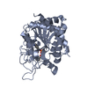
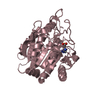
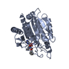
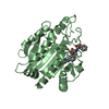
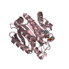
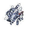
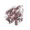
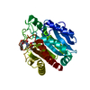
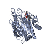
- PDB-2pja: CRYSTAL STRUCTURE OF ACTIVATED PORCINE PANCREATIC CARBOXYPEPTIDAS... -
+ Open data

- Basic information
| Entry | Database: PDB / ID: 2pja | ||||||
|---|---|---|---|---|---|---|---|
| Title | CRYSTAL STRUCTURE OF ACTIVATED PORCINE PANCREATIC CARBOXYPEPTIDASE B 3-{[(R)-1-((S)-2-Benzyloxycarbonylamino-3-phenyl-propionylamino)-2-methyl-propyl]-hydroxy-phosphinoyl}-2-(3-guanidino-phenyl)-propionic acid COMPLEX | ||||||
 Components Components | Carboxypeptidase B | ||||||
 Keywords Keywords | HYDROLASE / CARBOXYPEPTIDASE B / EXOPEPTIDASE / Phosphinate BASED INHIBITOR | ||||||
| Function / homology |  Function and homology information Function and homology informationcarboxypeptidase B / metallocarboxypeptidase activity / cytoplasmic vesicle / proteolysis / : / zinc ion binding Similarity search - Function | ||||||
| Biological species |  | ||||||
| Method |  X-RAY DIFFRACTION / SYNCHROTRON / FOURIER SYNTHESIS / Resolution: 1.7 Å X-RAY DIFFRACTION / SYNCHROTRON / FOURIER SYNTHESIS / Resolution: 1.7 Å | ||||||
 Authors Authors | Adler, M. / Whitlow, M. | ||||||
 Citation Citation | Journal: Acta Crystallogr.,Sect.D / Year: 2008 Title: Structures of potent selective peptide mimetics bound to carboxypeptidase B. Authors: Adler, M. / Buckman, B. / Bryant, J. / Chang, Z. / Chu, K. / Emayan, K. / Hrvatin, P. / Islam, I. / Morser, J. / Sukovich, D. / West, C. / Yuan, S. / Whitlow, M. #1: Journal: Biochemistry / Year: 2005Title: Crystal Structures of Potent Thiol-Based Inhibitors Bound to Carboxypeptidase B. Authors: Adler, M. / Bryant, J. / Buckman, B. / Islam, I. / Larsen, B. / Finster, S. / Kent, L. / May, K. / Mohan, R. / Yuan, S. / Whitlow, M. #2: Journal: Bioorg.Med.Chem.Lett. / Year: 2007Title: 3-Mercaptopropionic acids as efficacious inhibitors of activated thrombin activatable fibrinolysis inhibitor (TAFIa). Authors: Islam, I. / Bryant, J. / May, K. / Mohan, R. / Yuan, S. / Kent, L. / Morser, J. / Zhao, L. / Vergona, R. / White, K. / Adler, M. / Whitlow, M. / Buckman, B.O. | ||||||
| History |
|
- Structure visualization
| Structure viewer | Molecule: MolmilJmol/JSmol |
|---|
- Downloads & links
-Download
| PDBx/mmCIF format | 2pja.cif.gz | 207 KB | Display | PDBx/mmCIF format |
|---|---|---|---|---|
| PDB format | pdb2pja.ent.gz | 164.9 KB | Display | PDB format |
| PDBx/mmJSON format | 2pja.json.gz | Tree view | PDBx/mmJSON format | |
| Others |  Other downloads Other downloads |
-Validation report
| Arichive directory | https://data.pdbj.org/pub/pdb/validation_reports/pj/2pjaftp://data.pdbj.org/pub/pdb/validation_reports/pj/2pja | HTTPS FTP |
|---|
-Related structure data
| Related structure data |  2piyC  2pizC  2pj0C  2pj1C  2pj2C  2pj3C  2pj4C  2pj5C  2pj6C  2pj7C  2pj8C  2pj9C  2pjbC  2pjcC  1z5rS S: Starting model for refinement C: citing same article ( |
|---|---|
| Similar structure data |


-Links
 PDBj
PDBj
- Assembly
| Deposited unit | 
| ||||||||
|---|---|---|---|---|---|---|---|---|---|
| 1 | 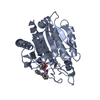
| ||||||||
| 2 | 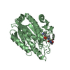
| ||||||||
| 3 | 
| ||||||||
| Unit cell |
|
-Components
| #1: Protein | Mass: 34739.859 Da / Num. of mol.: 3 / Fragment: CATALYTIC DOMAIN / Source method: isolated from a natural source / Source: (natural) #2: Chemical |   Mass: 65.409 Da / Num. of mol.: 3 / Source method: obtained synthetically / Formula: Zn Mass: 65.409 Da / Num. of mol.: 3 / Source method: obtained synthetically / Formula: Zn#3: Chemical | 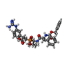  Mass: 623.636 Da / Num. of mol.: 3 / Source method: obtained synthetically / Formula: C31H38N5O7P Mass: 623.636 Da / Num. of mol.: 3 / Source method: obtained synthetically / Formula: C31H38N5O7P#4: Water | ChemComp-HOH / |  Mass: 18.015 Da / Num. of mol.: 624 / Source method: isolated from a natural source / Formula: H2O Mass: 18.015 Da / Num. of mol.: 624 / Source method: isolated from a natural source / Formula: H2OHas protein modification | Y | |
|---|
-Experimental details
-Experiment
| Experiment | Method: X-RAY DIFFRACTION / Number of used crystals: 1 |
|---|
- Sample preparation
| Crystal | Density Matthews: 2.1 Å3/Da / Density % sol: 41.41 % |
|---|---|
| Crystal grow | Temperature: 298 K / Method: vapor diffusion, hanging drop / pH: 6.5 Details: SODIUM CHLORIDE, TRIS, SODIUM CACODYLATE, PEG 8000, pH 6.50, VAPOR DIFFUSION, HANGING DROP, temperature 298K |
-Data collection
| Diffraction | Mean temperature: 100 K |
|---|---|
| Diffraction source | Source: SYNCHROTRON / Site: SSRL  / Beamline: BL7-1 / Wavelength: 1.08 / Beamline: BL7-1 / Wavelength: 1.08 |
| Detector | Type: MAR scanner 345 mm plate / Detector: IMAGE PLATE / Date: Apr 24, 2001 / Details: SI(111) |
| Radiation | Monochromator: SI(111) / Protocol: SINGLE WAVELENGTH / Monochromatic (M) / Laue (L): M / Scattering type: x-ray |
| Radiation wavelength | Wavelength: 1.08 Å / Relative weight: 1 |
| Reflection | Resolution: 1.7→21.4 Å / Num. obs: 93217 / % possible obs: 96.2 % / Redundancy: 3.5 % / Biso Wilson estimate: 17.6 Å2 / Rsym value: 0.087 / Net I/σ(I): 7.2 |
| Reflection shell | Resolution: 1.7→1.74 Å / Redundancy: 3.4 % / Mean I/σ(I) obs: 2.2 / Rsym value: 0.353 / % possible all: 89.1 |
- Processing
| Software |
| ||||||||||||||||||||||||||||||||||||||||||||||||||||||||||||||||||||||||||||||||
|---|---|---|---|---|---|---|---|---|---|---|---|---|---|---|---|---|---|---|---|---|---|---|---|---|---|---|---|---|---|---|---|---|---|---|---|---|---|---|---|---|---|---|---|---|---|---|---|---|---|---|---|---|---|---|---|---|---|---|---|---|---|---|---|---|---|---|---|---|---|---|---|---|---|---|---|---|---|---|---|---|---|
| Refinement | Method to determine structure: FOURIER SYNTHESIS Starting model: PDB ENTRY 1Z5R Resolution: 1.7→21.4 Å / Rfactor Rfree error: 0.004 / Data cutoff high absF: 2421454.56 / Data cutoff low absF: 0 / Isotropic thermal model: RESTRAINED / Cross valid method: THROUGHOUT / σ(F): 2 / Stereochemistry target values: Engh & Huber
| ||||||||||||||||||||||||||||||||||||||||||||||||||||||||||||||||||||||||||||||||
| Solvent computation | Solvent model: FLAT MODEL / Bsol: 45.4 Å2 / ksol: 0.36 e/Å3 | ||||||||||||||||||||||||||||||||||||||||||||||||||||||||||||||||||||||||||||||||
| Displacement parameters | Biso mean: 21.3 Å2
| ||||||||||||||||||||||||||||||||||||||||||||||||||||||||||||||||||||||||||||||||
| Refine analyze |
| ||||||||||||||||||||||||||||||||||||||||||||||||||||||||||||||||||||||||||||||||
| Refinement step | Cycle: LAST / Resolution: 1.7→21.4 Å
| ||||||||||||||||||||||||||||||||||||||||||||||||||||||||||||||||||||||||||||||||
| Refine LS restraints |
| ||||||||||||||||||||||||||||||||||||||||||||||||||||||||||||||||||||||||||||||||
| LS refinement shell | Resolution: 1.7→1.76 Å / Rfactor Rfree error: 0.015 / Total num. of bins used: 10
| ||||||||||||||||||||||||||||||||||||||||||||||||||||||||||||||||||||||||||||||||
| Xplor file |
|
