[English] 日本語
Yorodumi
- PDB-1zg7: Crystal Structure of 2-(5-{[amino(imino)methyl]amino}-2-chlorophe... -

+
Open data


ID or keywords:

Loading...

-
Basic information

Entry
Database: PDB / ID: 1zg7
Title
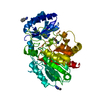
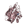
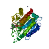
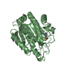
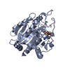
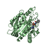
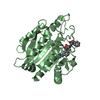
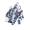
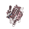
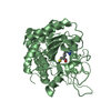
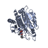
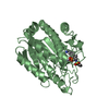
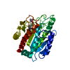
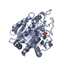
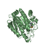
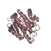
Crystal Structure of 2-(5-{[amino(imino)methyl]amino}-2-chlorophenyl)-3-sulfanylpropanoic acid Bound to Activated Porcine Pancreatic Carboxypeptidase B
Componentsprocarboxypeptidase B
KeywordsHYDROLASE / CARBOXYPEPTIDASE B / EXOPEPTIDASE / thiol based inhibitor
Function / homology
Function and homology information


carboxypeptidase B / metallocarboxypeptidase activity / cytoplasmic vesicle / proteolysis / extracellular space / zinc ion binding
Similarity search - Function
Carboxypeptidase B, carboxypeptidase domain / Carboxypeptidase, activation peptide / Metallocarboxypeptidase-like, propeptide / Carboxypeptidase activation peptide / Zinc carboxypeptidases, zinc-binding region 2 signature. / Zinc carboxypeptidases, zinc-binding region 1 signature. / Peptidase family M14 domain profile. / Zn_pept / Peptidase M14, carboxypeptidase A / Zinc carboxypeptidase ...Carboxypeptidase B, carboxypeptidase domain / Carboxypeptidase, activation peptide / Metallocarboxypeptidase-like, propeptide / Carboxypeptidase activation peptide / Zinc carboxypeptidases, zinc-binding region 2 signature. / Zinc carboxypeptidases, zinc-binding region 1 signature. / Peptidase family M14 domain profile. / Zn_pept / Peptidase M14, carboxypeptidase A / Zinc carboxypeptidase / Zn peptidases / Aminopeptidase / 3-Layer(aba) Sandwich / Alpha Beta
Similarity search - Domain/homology
Chem-P20 / Carboxypeptidase B
Similarity search - Component
Biological speciesSus scrofa (pig)
MethodX-RAY DIFFRACTION / SYNCHROTRON / MOLECULAR REPLACEMENT / Resolution: 1.75 Å
AuthorsAdler, M. / Bryant, J. / Buckman, B. / Islam, I. / Larsen, B. / Finster, S. / Kent, L. / May, K. / Mohan, R. / Yuan, S. / Whitlow, M.
CitationJournal: Biochemistry / Year: 2005
Title: Crystal structures of potent thiol-based inhibitors bound to carboxypeptidase b.
Authors: Adler, M. / Bryant, J. / Buckman, B. / Islam, I. / Larsen, B. / Finster, S. / Kent, L. / May, K. / Mohan, R. / Yuan, S. / Whitlow, M.
History
DepositionApr 20, 2005Deposition site: RCSB / Processing site: RCSB
Revision 1.0Jul 12, 2005Provider: repository / Type: Initial release
Revision 1.1Apr 30, 2008Group: Version format compliance
Revision 1.2Jul 13, 2011Group: Version format compliance
Revision 1.3Aug 23, 2023Group: Data collection / Database references ...Data collection / Database references / Derived calculations / Refinement description
Category: chem_comp_atom / chem_comp_bond ...chem_comp_atom / chem_comp_bond / database_2 / pdbx_initial_refinement_model / pdbx_struct_conn_angle / struct_conn / struct_site
Item: _database_2.pdbx_DOI / _database_2.pdbx_database_accession ..._database_2.pdbx_DOI / _database_2.pdbx_database_accession / _pdbx_struct_conn_angle.ptnr1_auth_comp_id / _pdbx_struct_conn_angle.ptnr1_auth_seq_id / _pdbx_struct_conn_angle.ptnr1_label_asym_id / _pdbx_struct_conn_angle.ptnr1_label_atom_id / _pdbx_struct_conn_angle.ptnr1_label_comp_id / _pdbx_struct_conn_angle.ptnr1_label_seq_id / _pdbx_struct_conn_angle.ptnr3_auth_comp_id / _pdbx_struct_conn_angle.ptnr3_auth_seq_id / _pdbx_struct_conn_angle.ptnr3_label_asym_id / _pdbx_struct_conn_angle.ptnr3_label_atom_id / _pdbx_struct_conn_angle.ptnr3_label_comp_id / _pdbx_struct_conn_angle.ptnr3_label_seq_id / _pdbx_struct_conn_angle.value / _struct_conn.pdbx_dist_value / _struct_conn.ptnr1_auth_comp_id / _struct_conn.ptnr1_auth_seq_id / _struct_conn.ptnr1_label_asym_id / _struct_conn.ptnr1_label_atom_id / _struct_conn.ptnr1_label_comp_id / _struct_conn.ptnr1_label_seq_id / _struct_conn.ptnr2_auth_comp_id / _struct_conn.ptnr2_auth_seq_id / _struct_conn.ptnr2_label_asym_id / _struct_conn.ptnr2_label_atom_id / _struct_conn.ptnr2_label_comp_id / _struct_conn.ptnr2_label_seq_id / _struct_site.pdbx_auth_asym_id / _struct_site.pdbx_auth_comp_id / _struct_site.pdbx_auth_seq_id
Remark 600HETEROGEN THE INHIBITION CONSTANT (KI APPARENT) OF 2-(5-{[AMINO (IMINO)METHYL] AMINO}-2- ...HETEROGEN THE INHIBITION CONSTANT (KI APPARENT) OF 2-(5-{[AMINO (IMINO)METHYL] AMINO}-2-CHLOROPHENYL)-3-SULFANYLPROPANOIC ACID WAS MEASURED AS 7 NM AGAINST PORCINE PANCREATIC CARBOXYPEPTIDASE B. THE ASSAY FOR INHIBITION OF PORCINE PANCREATIC CPB (SIGMA, ST. LOUIS, MO) WAS PERFORMED IN A 96 OR 384-WELL FORMAT ADAPTED FROM PUBLISHED PROTOCOLS. PURIFIED PP-CPB (2 NM) WAS INCUBATED WITH TEST COMPOUNDS IN 20 MM HEPES PH 7.4, 150 MM NACL, 5 MM CACL2 FOR 2 MINUTES PRIOR TO THE ADDITION OF THE 0.6 MM HIPPURYL-L -ARGININE SUBSTRATE. AFTER 30 MINUTES AT ROOM TEMPERATURE, THE AMOUNT OF SUBSTRATE HYDROLYZED WAS DETERMINED BY CONVERSION OF THE PRODUCT, HIPPURIC ACID, TO A CHROMOGEN WITH SODIUM PHOSPHATE BUFFER, PH 8.3 (F.C. 0.8 MM) AND CYANURIC CHLORIDE/DIOXANE (F.C. 0.9% W/V) UNDER CHEMICAL FUME HOOD. FOLLOWING CENTRIFUGATION OF THE MICROTITER PLATES AND TRANSFER OF THE SUPERNATANT TO A CLEAN PLATE, ABSORBANCE OF THE SUPERNATANT IS READ AT 382 NM. THE IC50 OF THE COMPOUND WAS DETERMINED USING THE 4-PARAMETER EQUATION FROM AN 8-POINT DOSE RESPONSE CURVE, EACH COMPOUND TESTED IN DUPLICATE.

-
Structure visualization

Structure viewerMolecule:
MolmilJmol/JSmol

Downloads & links

-
Assembly

Deposited unit
A: procarboxypeptidase B
B: procarboxypeptidase B
C: procarboxypeptidase B
hetero molecules


Theoretical massNumber of molelcules
Total (without water)105,2379
Polymers104,2203
Non-polymers1,0176
Water11,620645
1
A: procarboxypeptidase B
hetero molecules


Theoretical massNumber of molelcules
Total (without water)35,0793
Polymers34,7401
Non-polymers3392
Water181
TypeNameSymmetry operationNumber
identity operation1_555x,y,z1
2
B: procarboxypeptidase B
hetero molecules


Theoretical massNumber of molelcules
Total (without water)35,0793
Polymers34,7401
Non-polymers3392
Water181
TypeNameSymmetry operationNumber
identity operation1_555x,y,z1
3
C: procarboxypeptidase B
hetero molecules


Theoretical massNumber of molelcules
Total (without water)35,0793
Polymers34,7401
Non-polymers3392
Water181
TypeNameSymmetry operationNumber
identity operation1_555x,y,z1
Unit cell
Length a, b, c (Å)67.504, 100.369, 136.004
Angle α, β, γ (deg.)90.00, 90.00, 90.00
Int Tables number19
Space group name H-MP212121
DetailsThe biological assembly is a monomer that can be generated from either chain A, chain B, or chain C.

-
Components

#1: Protein procarboxypeptidase B


Mass: 34739.859 Da / Num. of mol.: 3 / Fragment: Catalytic Domain / Source method: isolated from a natural source / Source: (natural) Sus scrofa (pig) / References: UniProt: P09955, carboxypeptidase B
#2: Chemical ChemComp-ZN / ZINC ION


Mass: 65.409 Da / Num. of mol.: 3 / Source method: obtained synthetically / Formula: Zn
#3: Chemical ChemComp-P20 / 2-(5-{[AMINO(IMINO)METHYL]AMINO}-2-CHLOROPHENYL)-3-SULFANYLPROPANOIC ACID


Mass: 273.739 Da / Num. of mol.: 3 / Source method: obtained synthetically / Formula: C10H12ClN3O2S
#4: Water ChemComp-HOH / water


Mass: 18.015 Da / Num. of mol.: 645 / Source method: isolated from a natural source / Formula: H2O

-
Experimental details

-
Experiment

ExperimentMethod: X-RAY DIFFRACTION / Number of used crystals: 1

-
Sample preparation

CrystalDensity Matthews: 2.2 Å3/Da / Density % sol: 44.2 %
Crystal growTemperature: 298 K / Method: vapor diffusion, hanging drop / pH: 6.5
Details: sodium chloride, Tris, sodium cacodylate, PEG 8000, pH 6.50, VAPOR DIFFUSION, HANGING DROP, temperature 298K

-
Data collection

DiffractionMean temperature: 100 K
Diffraction sourceSource: SYNCHROTRON / Site: SSRL / Beamline: BL7-1 / Wavelength: 1.08 / Wavelength: 1.08 Å
DetectorType: MAR scanner 345 mm plate / Detector: IMAGE PLATE / Date: Dec 7, 2000
RadiationMonochromator: SI(111) / Protocol: SINGLE WAVELENGTH / Monochromatic (M) / Laue (L): M / Scattering type: x-ray
Radiation wavelength
IDWavelength (Å)Relative weight
11.081
21.081
ReflectionResolution: 1.75→20 Å / Num. obs: 93605 / % possible obs: 99.9 % / Observed criterion σ(F): 0 / Observed criterion σ(I): 0 / Redundancy: 3.3 % / Biso Wilson estimate: 24.9 Å2 / Rsym value: 0.068 / Net I/σ(I): 6.6
Reflection shellResolution: 1.75→1.83 Å / Redundancy: 3.3 % / Mean I/σ(I) obs: 1.7 / Num. unique all: 11051 / Rsym value: 0.36 / % possible all: 100

-
Processing

Software
NameVersionClassification
MOSFLMdata reduction
SCALAdata scaling
MOLREPphasing
X-PLOR3.1refinement
CCP4(SCALA)data scaling
RefinementMethod to determine structure: MOLECULAR REPLACEMENT
Starting model: PDB ENTRY 1NAS CATALYTIC DOMAIN
Resolution: 1.75→8 Å / Isotropic thermal model: OVERALL / σ(F): 2 / Stereochemistry target values: Engh & Huber
RfactorNum. reflection% reflectionSelection details
Rfree0.262 3473 -RANDOM
Rwork0.208 ---
all0.213 92660 --
obs0.211 90906 99.9 %-
Displacement parametersBiso mean: 29.25 Å2
Baniso -1Baniso -2Baniso -3
1--4.8686 Å20 Å20 Å2
2---4.6192 Å20 Å2
3---9.4878 Å2
Refinement stepCycle: LAST / Resolution: 1.75→8 Å
ProteinNucleic acidLigandSolventTotal
Num. atoms7320 0 54 645 8019
Refine LS restraints
Refine-IDTypeDev ideal
X-RAY DIFFRACTIONx_bond_d0.012
X-RAY DIFFRACTIONx_bond_d_na
X-RAY DIFFRACTIONx_bond_d_prot
X-RAY DIFFRACTIONx_angle_d
X-RAY DIFFRACTIONx_angle_d_na
X-RAY DIFFRACTIONx_angle_d_prot
X-RAY DIFFRACTIONx_angle_deg1.81
X-RAY DIFFRACTIONx_angle_deg_na
X-RAY DIFFRACTIONx_angle_deg_prot
X-RAY DIFFRACTIONx_dihedral_angle_d
X-RAY DIFFRACTIONx_dihedral_angle_d_na
X-RAY DIFFRACTIONx_dihedral_angle_d_prot
X-RAY DIFFRACTIONx_improper_angle_d1.42
X-RAY DIFFRACTIONx_improper_angle_d_na
X-RAY DIFFRACTIONx_improper_angle_d_prot
X-RAY DIFFRACTIONx_mcbond_it
X-RAY DIFFRACTIONx_mcangle_it
X-RAY DIFFRACTIONx_scbond_it
X-RAY DIFFRACTIONx_scangle_it
LS refinement shell

Refine-ID: X-RAY DIFFRACTION

Resolution (Å)Rfactor RfreeNum. reflection Rfree% reflection Rfree (%)Rfactor RworkNum. reflection RworkNum. reflection obs% reflection obs (%)
1.75-1.830.33773983.70.36551065311051100
1.83-1.920.3464130.323611107100
1.92-2.040.31164330.28711194100
2.04-2.20.29164210.257111276100
2.2-2.410.26784540.23711368100
2.41-2.750.24954490.215911440100
2.75-3.420.274540.191158699.9
3.42-80.23244510.15861188499.9

+
About Yorodumi

-
News

-
Feb 9, 2022. New format data for meta-information of EMDB entries

New format data for meta-information of EMDB entries

  • Version 3 of the EMDB header file is now the official format.
  • The previous official version 1.9 will be removed from the archive.

Related info.:EMDB header

External links:wwPDB to switch to version 3 of the EMDB data model

-
Aug 12, 2020. Covid-19 info

Covid-19 info

URL: https://pdbjlc1.pdbj.org/emnavi/covid19.php

New page: Covid-19 featured information page in EM Navigator.

Related info.:Covid-19 info / Mar 5, 2020. Novel coronavirus structure data

+
Mar 5, 2020. Novel coronavirus structure data

Novel coronavirus structure data

Related info.:Yorodumi Speices / Aug 12, 2020. Covid-19 info

External links:COVID-19 featured content - PDBj / Molecule of the Month (242):Coronavirus Proteases

+
Jan 31, 2019. EMDB accession codes are about to change! (news from PDBe EMDB page)

EMDB accession codes are about to change! (news from PDBe EMDB page)

  • The allocation of 4 digits for EMDB accession codes will soon come to an end. Whilst these codes will remain in use, new EMDB accession codes will include an additional digit and will expand incrementally as the available range of codes is exhausted. The current 4-digit format prefixed with “EMD-” (i.e. EMD-XXXX) will advance to a 5-digit format (i.e. EMD-XXXXX), and so on. It is currently estimated that the 4-digit codes will be depleted around Spring 2019, at which point the 5-digit format will come into force.
  • The EM Navigator/Yorodumi systems omit the EMD- prefix.

Related info.:Q: What is EMD? / ID/Accession-code notation in Yorodumi/EM Navigator

External links:EMDB Accession Codes are Changing Soon! / Contact to PDBj

+
Jul 12, 2017. Major update of PDB

Major update of PDB

  • wwPDB released updated PDB data conforming to the new PDBx/mmCIF dictionary.
  • This is a major update changing the version number from 4 to 5, and with Remediation, in which all the entries are updated.
  • In this update, many items about electron microscopy experimental information are reorganized (e.g. em_software).
  • Now, EM Navigator and Yorodumi are based on the updated data.

External links:wwPDB Remediation / Enriched Model Files Conforming to OneDep Data Standards Now Available in the PDB FTP Archive

-
Yorodumi

Thousand views of thousand structures

  • Yorodumi is a browser for structure data from EMDB, PDB, SASBDB, etc.
  • This page is also the successor to EM Navigator detail page, and also detail information page/front-end page for Omokage search.
  • The word "yorodu" (or yorozu) is an old Japanese word meaning "ten thousand". "mi" (miru) is to see.

Related info.:EMDB / PDB / SASBDB / Comparison of 3 databanks / Yorodumi Search / Aug 31, 2016. New EM Navigator & Yorodumi / Yorodumi Papers / Jmol/JSmol / Function and homology information / Changes in new EM Navigator and Yorodumi

Read more