 Movie
Movie Controller

[English] 日本語
 Yorodumi
- PDB-1kwm: Human procarboxypeptidase B: Three-dimensional structure and impl... -
+ Open data

- Basic information
| Entry | Database: PDB / ID: 1kwm | ||||||
|---|---|---|---|---|---|---|---|
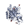
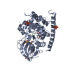
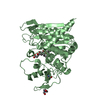
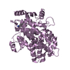
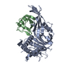
| Title | Human procarboxypeptidase B: Three-dimensional structure and implications for thrombin-activatable fibrinolysis inhibitor (TAFI) | ||||||
 Components Components | Procarboxypeptidase B | ||||||
 Keywords Keywords | HYDROLASE / Procarboxypeptidase B | ||||||
| Function / homology |  Function and homology information Function and homology informationcarboxypeptidase B / Developmental Lineage of Pancreatic Acinar Cells / Metabolism of Angiotensinogen to Angiotensins / carboxypeptidase activity / metallocarboxypeptidase activity / cytoplasmic vesicle / proteolysis / : / zinc ion binding Similarity search - Function | ||||||
| Biological species |  Homo sapiens (human) Homo sapiens (human) | ||||||
| Method |  X-RAY DIFFRACTION / SYNCHROTRON / MOLECULAR REPLACEMENT / Resolution: 1.6 Å X-RAY DIFFRACTION / SYNCHROTRON / MOLECULAR REPLACEMENT / Resolution: 1.6 Å | ||||||
 Authors Authors | Pereira, P.J.B. / Segura-Martin, S. / Ferrer-Orta, C. / Vendrell, J. / Aviles, F.-X. / Coll, M. / Gomis-Rueth, F.-X. | ||||||
 Citation Citation | Journal: J.Mol.Biol. / Year: 2002 Title: Human procarboxypeptidase B: three-dimensional structure and implications for thrombin-activatable fibrinolysis inhibitor (TAFI). Authors: Barbosa Pereira, P.J. / Segura-Martin, S. / Oliva, B. / Ferrer-Orta, C. / Aviles, F.X. / Coll, M. / Gomis-Ruth, F.X. / Vendrell, J. | ||||||
| History |
|
- Structure visualization
| Structure viewer | Molecule: MolmilJmol/JSmol |
|---|
- Downloads & links
-Download
| PDBx/mmCIF format | 1kwm.cif.gz | 193.4 KB | Display | PDBx/mmCIF format |
|---|---|---|---|---|
| PDB format | pdb1kwm.ent.gz | 150.6 KB | Display | PDB format |
| PDBx/mmJSON format | 1kwm.json.gz | Tree view | PDBx/mmJSON format | |
| Others |  Other downloads Other downloads |
-Validation report
| Arichive directory | https://data.pdbj.org/pub/pdb/validation_reports/kw/1kwmftp://data.pdbj.org/pub/pdb/validation_reports/kw/1kwm | HTTPS FTP |
|---|
-Related structure data
| Similar structure data |
|---|


-Links
 PDBj
PDBj


- Assembly
| Deposited unit | 
| ||||||||
|---|---|---|---|---|---|---|---|---|---|
| 1 | 
| ||||||||
| 2 | 
| ||||||||
| Unit cell |
|
-Components
| #1: Protein | Mass: 45955.488 Da / Num. of mol.: 2 Source method: isolated from a genetically manipulated source Source: (gene. exp.) Homo sapiens (human) / Plasmid: pPIC9 / Production host:  Pichia pastoris (fungus) / References: UniProt: P15086, carboxypeptidase B Pichia pastoris (fungus) / References: UniProt: P15086, carboxypeptidase B#2: Chemical |   Mass: 65.409 Da / Num. of mol.: 2 / Source method: obtained synthetically / Formula: Zn Mass: 65.409 Da / Num. of mol.: 2 / Source method: obtained synthetically / Formula: Zn#3: Chemical | ChemComp-CIT / |   Mass: 192.124 Da / Num. of mol.: 1 / Source method: obtained synthetically / Formula: C6H8O7 Mass: 192.124 Da / Num. of mol.: 1 / Source method: obtained synthetically / Formula: C6H8O7#4: Water | ChemComp-HOH / |  Mass: 18.015 Da / Num. of mol.: 814 / Source method: isolated from a natural source / Formula: H2O Mass: 18.015 Da / Num. of mol.: 814 / Source method: isolated from a natural source / Formula: H2OHas protein modification | Y | |
|---|
-Experimental details
-Experiment
| Experiment | Method: X-RAY DIFFRACTION / Number of used crystals: 1 |
|---|
- Sample preparation
| Crystal | Density Matthews: 2.46 Å3/Da / Density % sol: 49.93 % | |||||||||||||||||||||||||||||||||||||||||||||||||
|---|---|---|---|---|---|---|---|---|---|---|---|---|---|---|---|---|---|---|---|---|---|---|---|---|---|---|---|---|---|---|---|---|---|---|---|---|---|---|---|---|---|---|---|---|---|---|---|---|---|---|
| Crystal grow | Temperature: 293 K / Method: vapor diffusion, hanging drop / pH: 6 Details: MES, sodium citrate, sodium chloride, pH 6.0, VAPOR DIFFUSION, HANGING DROP, temperature 293K | |||||||||||||||||||||||||||||||||||||||||||||||||
| Crystal grow | *PLUS Temperature: 20 ℃ / pH: 7 | |||||||||||||||||||||||||||||||||||||||||||||||||
| Components of the solutions | *PLUS
|
-Data collection
| Diffraction | Mean temperature: 100 K |
|---|---|
| Diffraction source | Source: SYNCHROTRON / Site: EMBL/DESY, HAMBURG  / Beamline: BW7A / Wavelength: 0.97935 Å / Beamline: BW7A / Wavelength: 0.97935 Å |
| Detector | Type: MARRESEARCH / Detector: IMAGE PLATE / Date: Dec 15, 1999 |
| Radiation | Protocol: SINGLE WAVELENGTH / Monochromatic (M) / Laue (L): M / Scattering type: x-ray |
| Radiation wavelength | Wavelength: 0.97935 Å / Relative weight: 1 |
| Reflection | Resolution: 1.6→27.4 Å / Num. all: 104805 / Num. obs: 104805 / % possible obs: 89.7 % / Observed criterion σ(F): 0 / Observed criterion σ(I): 0 / Rsym value: 0.05 / Net I/σ(I): 8.4 |
| Reflection shell | Resolution: 1.6→1.68 Å / Mean I/σ(I) obs: 2.7 / Rsym value: 0.241 / % possible all: 81.9 |
| Reflection | *PLUS Highest resolution: 1.6 Å / Redundancy: 2.9 % / Num. measured all: 303465 / Rmerge(I) obs: 0.05 |
| Reflection shell | *PLUS % possible obs: 81.9 % / Redundancy: 2.4 % / Rmerge(I) obs: 0.241 |
- Processing
| Software |
| ||||||||||||||||||||||||||||||
|---|---|---|---|---|---|---|---|---|---|---|---|---|---|---|---|---|---|---|---|---|---|---|---|---|---|---|---|---|---|---|---|
| Refinement | Method to determine structure: MOLECULAR REPLACEMENT / Resolution: 1.6→27.4 Å / Cross valid method: THROUGHOUT / σ(F): 0 / σ(I): 0 / Stereochemistry target values: Engh & Huber
| ||||||||||||||||||||||||||||||
| Refinement step | Cycle: LAST / Resolution: 1.6→27.4 Å
| ||||||||||||||||||||||||||||||
| Software | *PLUS Name: SHELXL / Version: 97 / Classification: refinement | ||||||||||||||||||||||||||||||
| Refinement | *PLUS Highest resolution: 1.6 Å / % reflection Rfree: 5 % / Rfactor Rwork: 0.136 | ||||||||||||||||||||||||||||||
| Solvent computation | *PLUS | ||||||||||||||||||||||||||||||
| Displacement parameters | *PLUS | ||||||||||||||||||||||||||||||
| Refine LS restraints | *PLUS
|
