[English] 日本語
Yorodumi
- PDB-5lyf: Crystal structure of 1 in complex with tafCPB -

+
Open data


ID or keywords:

Loading...

-
Basic information

Entry
Database: PDB / ID: 5lyf
TitleCrystal structure of 1 in complex with tafCPB
ComponentsCarboxypeptidase B
KeywordsHYDROLASE / tafinized carboxypeptidase B / HYRDROLASE
Function / homology
Function and homology information


carboxypeptidase B / metallocarboxypeptidase activity / cytoplasmic vesicle / proteolysis / extracellular space / zinc ion binding
Similarity search - Function
Carboxypeptidase B, carboxypeptidase domain / Carboxypeptidase, activation peptide / Metallocarboxypeptidase-like, propeptide / Carboxypeptidase activation peptide / Zinc carboxypeptidases, zinc-binding region 2 signature. / Zinc carboxypeptidases, zinc-binding region 1 signature. / Peptidase family M14 domain profile. / Zn_pept / Peptidase M14, carboxypeptidase A / Zinc carboxypeptidase ...Carboxypeptidase B, carboxypeptidase domain / Carboxypeptidase, activation peptide / Metallocarboxypeptidase-like, propeptide / Carboxypeptidase activation peptide / Zinc carboxypeptidases, zinc-binding region 2 signature. / Zinc carboxypeptidases, zinc-binding region 1 signature. / Peptidase family M14 domain profile. / Zn_pept / Peptidase M14, carboxypeptidase A / Zinc carboxypeptidase / Zn peptidases / Aminopeptidase / 3-Layer(aba) Sandwich / Alpha Beta
Similarity search - Domain/homology
Chem-7B6 / Carboxypeptidase B
Similarity search - Component
Biological speciesSus scrofa (pig)
MethodX-RAY DIFFRACTION / SYNCHROTRON / Resolution: 2.01 Å
AuthorsSchreuder, H. / Liesum, A. / Loenze, P.
CitationJournal: J. Med. Chem. / Year: 2016
Title: Sulfamide as Zinc Binding Motif in Small Molecule Inhibitors of Activated Thrombin Activatable Fibrinolysis Inhibitor (TAFIa).
Authors: Halland, N. / Czech, J. / Czechtizky, W. / Evers, A. / Follmann, M. / Kohlmann, M. / Schreuder, H.A. / Kallus, C.
History
DepositionSep 28, 2016Deposition site: PDBE / Processing site: PDBE
Revision 1.0Oct 26, 2016Provider: repository / Type: Initial release
Revision 1.1Dec 21, 2016Group: Database references

-
Structure visualization

Structure viewerMolecule:
MolmilJmol/JSmol

Downloads & links

-
Assembly

Deposited unit
A: Carboxypeptidase B
hetero molecules


Theoretical massNumber of molelcules
Total (without water)35,4935
Polymers34,8601
Non-polymers6334
Water10,485582
1


  • Idetical with deposited unit
  • defined by software
TypeNameSymmetry operationNumber
identity operation1_555x,y,z1
Buried area150 Å2
ΔGint-36 kcal/mol
Surface area11900 Å2
MethodPISA
Unit cell
Length a, b, c (Å)81.490, 81.490, 95.734
Angle α, β, γ (deg.)90.00, 90.00, 90.00
Int Tables number92
Space group name H-MP41212
Components on special symmetry positions
IDModelComponents
11A-923-

HOH

21A-950-

HOH

-
Components

#1: Protein Carboxypeptidase B /


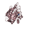
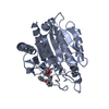
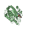
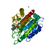
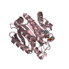
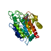
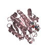
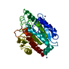
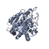
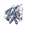
Mass: 34859.969 Da / Num. of mol.: 1
Source method: isolated from a genetically manipulated source
Source: (gene. exp.) Sus scrofa (pig) / Gene: CPB1, CPB / Production host: Komagataella pastoris CBS 7435 (fungus) / References: UniProt: P09955, carboxypeptidase B
#2: Chemical ChemComp-ZN / ZINC ION


Mass: 65.409 Da / Num. of mol.: 3 / Source method: obtained synthetically / Formula: Zn
#3: Chemical ChemComp-7B6 / (2~{S})-6-azanyl-2-[[(2~{R})-1-[[(1~{R},2~{S},4~{S})-2-bicyclo[2.2.1]heptanyl]amino]-3-cyclohexyl-1-oxidanylidene-propan-2-yl]carbamoylamino]hexanoic acid


Mass: 436.588 Da / Num. of mol.: 1 / Source method: obtained synthetically / Formula: C23H40N4O4
#4: Water ChemComp-HOH / water / Water


Mass: 18.015 Da / Num. of mol.: 582 / Source method: isolated from a natural source / Formula: H2O

-
Experimental details

-
Experiment

ExperimentMethod: X-RAY DIFFRACTION / Number of used crystals: 1

-
Sample preparation

CrystalDensity Matthews: 2.3 Å3/Da / Density % sol: 46.5 %
Crystal growTemperature: 293 K / Method: vapor diffusion, hanging drop
Details: THE PURIFIED PROTEIN WAS DISSOLVED IN 50 MM TRIS-HCL, PH 7.5 AND CONCENTRATED TO 11 MG/ML. 1 UL OF PROTEIN SOLUTION WAS EQUILIBRATED AGAINST 1 UL OF RESERVOIR SOLUTIONS CONTAINING 16-20% ...Details: THE PURIFIED PROTEIN WAS DISSOLVED IN 50 MM TRIS-HCL, PH 7.5 AND CONCENTRATED TO 11 MG/ML. 1 UL OF PROTEIN SOLUTION WAS EQUILIBRATED AGAINST 1 UL OF RESERVOIR SOLUTIONS CONTAINING 16-20% PEG3350, 100 MM MES PH 5.5 AND 50 MM ZNACETATE

-
Data collection

DiffractionMean temperature: 100 K
Diffraction sourceSource: SYNCHROTRON / Site: ESRF / Beamline: ID14-1 / Wavelength: 0.934 Å
DetectorType: ADSC QUANTUM 210 / Detector: CCD / Date: Jan 29, 2007
RadiationProtocol: SINGLE WAVELENGTH / Monochromatic (M) / Laue (L): M / Scattering type: x-ray
Radiation wavelengthWavelength: 0.934 Å / Relative weight: 1
ReflectionResolution: 2.01→81.42 Å / Num. obs: 21825 / % possible obs: 98.7 % / Observed criterion σ(F): -3 / Redundancy: 10.4 % / Biso Wilson estimate: 23.4 Å2 / Rmerge(I) obs: 0.113 / Net I/σ(I): 15.7
Reflection shellResolution: 2.01→2.1 Å / Redundancy: 9.4 % / Rmerge(I) obs: 0.284 / Mean I/σ(I) obs: 7.99 / % possible all: 90.7

-
Processing

Software
NameVersionClassification
REFMAC5.2.0019refinement
XDSdata reduction
XSCALEdata scaling
REFMAC5.2.0019phasing
RefinementResolution: 2.01→49.39 Å / Cor.coef. Fo:Fc: 0.939 / Cor.coef. Fo:Fc free: 0.845 / SU B: 6.973 / SU ML: 0.193 / Cross valid method: THROUGHOUT / ESU R: 0.234 / ESU R Free: 0.245 / Details: HYDROGENS HAVE BEEN ADDED IN THE RIDING POSITIONS
RfactorNum. reflection% reflectionSelection details
Rfree0.30479 1522 7 %RANDOM
Rwork0.18355 ---
obs0.19202 20300 99.15 %-
Solvent computationIon probe radii: 0.8 Å / Shrinkage radii: 0.8 Å / VDW probe radii: 1.4 Å
Displacement parametersBiso mean: 21.074 Å2
Baniso -1Baniso -2Baniso -3
1--0.13 Å20 Å20 Å2
2---0.13 Å20 Å2
3---0.25 Å2
Refinement stepCycle: 1 / Resolution: 2.01→49.39 Å
ProteinNucleic acidLigandSolventTotal
Num. atoms2446 0 34 582 3062
Refine LS restraints
Refine-IDTypeDev idealDev ideal targetNumber
X-RAY DIFFRACTIONr_bond_refined_d0.0220.0222552
X-RAY DIFFRACTIONr_bond_other_d
X-RAY DIFFRACTIONr_angle_refined_deg1.9561.9453476
X-RAY DIFFRACTIONr_angle_other_deg
X-RAY DIFFRACTIONr_dihedral_angle_1_deg7.2525304
X-RAY DIFFRACTIONr_dihedral_angle_2_deg33.3524.034119
X-RAY DIFFRACTIONr_dihedral_angle_3_deg16.96515395
X-RAY DIFFRACTIONr_dihedral_angle_4_deg16.6511510
X-RAY DIFFRACTIONr_chiral_restr0.1510.2369
X-RAY DIFFRACTIONr_gen_planes_refined0.010.021964
X-RAY DIFFRACTIONr_gen_planes_other
X-RAY DIFFRACTIONr_nbd_refined0.2520.21471
X-RAY DIFFRACTIONr_nbd_other
X-RAY DIFFRACTIONr_nbtor_refined0.3150.21719
X-RAY DIFFRACTIONr_nbtor_other
X-RAY DIFFRACTIONr_xyhbond_nbd_refined0.2670.2420
X-RAY DIFFRACTIONr_xyhbond_nbd_other
X-RAY DIFFRACTIONr_metal_ion_refined0.0570.21
X-RAY DIFFRACTIONr_metal_ion_other
X-RAY DIFFRACTIONr_symmetry_vdw_refined0.3110.242
X-RAY DIFFRACTIONr_symmetry_vdw_other
X-RAY DIFFRACTIONr_symmetry_hbond_refined0.2780.243
X-RAY DIFFRACTIONr_symmetry_hbond_other
X-RAY DIFFRACTIONr_symmetry_metal_ion_refined0.2720.22
X-RAY DIFFRACTIONr_symmetry_metal_ion_other
X-RAY DIFFRACTIONr_mcbond_it1.0961.51560
X-RAY DIFFRACTIONr_mcbond_other
X-RAY DIFFRACTIONr_mcangle_it1.66322439
X-RAY DIFFRACTIONr_mcangle_other
X-RAY DIFFRACTIONr_scbond_it2.59131191
X-RAY DIFFRACTIONr_scbond_other
X-RAY DIFFRACTIONr_scangle_it3.5124.51037
X-RAY DIFFRACTIONr_scangle_other
X-RAY DIFFRACTIONr_long_range_B_refined
X-RAY DIFFRACTIONr_long_range_B_other
X-RAY DIFFRACTIONr_rigid_bond_restr
X-RAY DIFFRACTIONr_sphericity_free
X-RAY DIFFRACTIONr_sphericity_bonded
LS refinement shellResolution: 2.013→2.065 Å / Total num. of bins used: 20
RfactorNum. reflection% reflection
Rfree0.386 83 -
Rwork0.238 1354 -
obs--90.21 %

+
About Yorodumi

-
News

-
Feb 9, 2022. New format data for meta-information of EMDB entries

New format data for meta-information of EMDB entries

  • Version 3 of the EMDB header file is now the official format.
  • The previous official version 1.9 will be removed from the archive.

Related info.:EMDB header

External links:wwPDB to switch to version 3 of the EMDB data model

-
Aug 12, 2020. Covid-19 info

Covid-19 info

URL: https://pdbj.org/emnavi/covid19.php

New page: Covid-19 featured information page in EM Navigator.

Related info.:Covid-19 info / Mar 5, 2020. Novel coronavirus structure data

+
Mar 5, 2020. Novel coronavirus structure data

Novel coronavirus structure data

Related info.:Yorodumi Speices / Aug 12, 2020. Covid-19 info

External links:COVID-19 featured content - PDBj / Molecule of the Month (242):Coronavirus Proteases

+
Jan 31, 2019. EMDB accession codes are about to change! (news from PDBe EMDB page)

EMDB accession codes are about to change! (news from PDBe EMDB page)

  • The allocation of 4 digits for EMDB accession codes will soon come to an end. Whilst these codes will remain in use, new EMDB accession codes will include an additional digit and will expand incrementally as the available range of codes is exhausted. The current 4-digit format prefixed with “EMD-” (i.e. EMD-XXXX) will advance to a 5-digit format (i.e. EMD-XXXXX), and so on. It is currently estimated that the 4-digit codes will be depleted around Spring 2019, at which point the 5-digit format will come into force.
  • The EM Navigator/Yorodumi systems omit the EMD- prefix.

Related info.:Q: What is EMD? / ID/Accession-code notation in Yorodumi/EM Navigator

External links:EMDB Accession Codes are Changing Soon! / Contact to PDBj

+
Jul 12, 2017. Major update of PDB

Major update of PDB

  • wwPDB released updated PDB data conforming to the new PDBx/mmCIF dictionary.
  • This is a major update changing the version number from 4 to 5, and with Remediation, in which all the entries are updated.
  • In this update, many items about electron microscopy experimental information are reorganized (e.g. em_software).
  • Now, EM Navigator and Yorodumi are based on the updated data.

External links:wwPDB Remediation / Enriched Model Files Conforming to OneDep Data Standards Now Available in the PDB FTP Archive

-
Yorodumi

Thousand views of thousand structures

  • Yorodumi is a browser for structure data from EMDB, PDB, SASBDB, etc.
  • This page is also the successor to EM Navigator detail page, and also detail information page/front-end page for Omokage search.
  • The word "yorodu" (or yorozu) is an old Japanese word meaning "ten thousand". "mi" (miru) is to see.

Related info.:EMDB / PDB / SASBDB / Comparison of 3 databanks / Yorodumi Search / Aug 31, 2016. New EM Navigator & Yorodumi / Yorodumi Papers / Jmol/JSmol / Function and homology information / Changes in new EM Navigator and Yorodumi

Read more