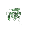
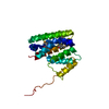
Entry Database : PDB / ID : 2wo1Title Crystal Structure of the EphA4 Ligand Binding Domain EPHRIN TYPE-A RECEPTOR Keywords / / / / / Function / homology Function Domain/homology Component
/ / / / / / / / / / / / / / / / / / / / / / / / / / / / / / / / / / / / / / / / / / / / / / / / / / / / / / / / / / / / / / / / / / / / / / / / / / / / / / / / / / / / / / / / / / / / / / / / / / / / / / / / / / / / / / / / / / / / / / / / / / / / / / / / / / / / / / / / / / / Biological species Homo sapiens (human)Method / / / Resolution : 1.85 Å Authors Bowden, T.A. / Aricescu, A.R. / Nettleship, J.E. / Siebold, C. / Rahman-Huq, N. / Owens, R.J. / Stuart, D.I. / Jones, E.Y. Journal : Structure / Year : 2009Title : Structural Plasticity of Eph-Receptor A4 Facilitates Cross-Class Ephrin SignallingAuthors : Bowden, T.A. / Aricescu, A.R. / Nettleship, J.E. / Siebold, C. / Rahman-Huq, N. / Owens, R.J. / Stuart, D.I. / Jones, E.Y. History Deposition Jul 21, 2009 Deposition site / Processing site Revision 1.0 Oct 27, 2009 Provider / Type Revision 1.1 Jul 13, 2011 Group / Version format complianceRevision 1.2 Nov 23, 2011 Group Revision 1.3 Feb 28, 2018 Group / Category Item _entity_src_gen.gene_src_common_name / _entity_src_gen.host_org_common_name ... _entity_src_gen.gene_src_common_name / _entity_src_gen.host_org_common_name / _entity_src_gen.pdbx_gene_src_scientific_name / _entity_src_gen.pdbx_host_org_cell_line / _entity_src_gen.pdbx_host_org_scientific_name / _entity_src_gen.pdbx_host_org_strain Revision 1.4 Dec 20, 2023 Group Data collection / Database references ... Data collection / Database references / Derived calculations / Other / Refinement description Category chem_comp_atom / chem_comp_bond ... chem_comp_atom / chem_comp_bond / database_2 / pdbx_database_status / pdbx_initial_refinement_model / struct_site Item _database_2.pdbx_DOI / _database_2.pdbx_database_accession ... _database_2.pdbx_DOI / _database_2.pdbx_database_accession / _pdbx_database_status.status_code_sf / _struct_site.pdbx_auth_asym_id / _struct_site.pdbx_auth_comp_id / _struct_site.pdbx_auth_seq_id Revision 1.5 Oct 9, 2024 Group / Category / pdbx_modification_feature
Show all Show less Remark 700 SHEET THE SHEET STRUCTURE OF THIS MOLECULE IS BIFURCATED. IN ORDER TO REPRESENT THIS FEATURE IN ... SHEET THE SHEET STRUCTURE OF THIS MOLECULE IS BIFURCATED. IN ORDER TO REPRESENT THIS FEATURE IN THE SHEET RECORDS BELOW, TWO SHEETS ARE DEFINED.
 Movie
Movie Controller


 Open data
 Basic information
 Components
 Keywords
 Function and homology information
 Homo sapiens (human)
 X-RAY DIFFRACTION /
  Authors
 Citation
 Structure visualization
 Downloads & links
 Other downloads


 PDBj
PDBj


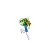
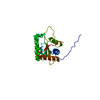
 Assembly
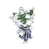


 Mass: 60.095 Da / Num. of mol.: 5 / Source method: obtained synthetically / Formula: C3H8O
 Mass: 18.015 Da / Num. of mol.: 297 / Source method: isolated from a natural source / Formula: H2O
 Sample preparation
 / Beamline: BM14 / Wavelength: 0.977
  Processing