- PDB-3u66: Crystal structure of T6SS SciP/TssL from Escherichia Coli Enteroa... -
+
Open data
ID or keywords:
Loading...
-
Basic information
Entry
Database: PDB / ID: 3u66
Title
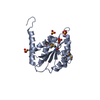
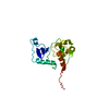
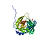
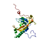
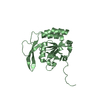
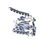
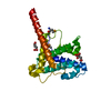
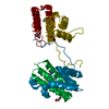
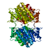
Crystal structure of T6SS SciP/TssL from Escherichia Coli Enteroaggregative 042
Components
Putative type VI secretion protein
Keywords
UNKNOWN FUNCTION / Duble helical bundle / Protein secretion / attached to inner membrane
Function / homology
Type IV / VI secretion system, DotU / Type IV / VI secretion system, DotU / Type IV / VI secretion system, DotU superfamily / Type VI secretion system protein DotU / Serine Threonine Protein Phosphatase 5, Tetratricopeptide repeat / Alpha Horseshoe / Mainly Alpha / Type VI secretion protein
Function and homology information
Biological species
Escherichia coli (E. coli)
Method
X-RAY DIFFRACTION / SYNCHROTRON / SAD / Resolution: 2.63 Å
Mass: 18.015 Da / Num. of mol.: 31 / Source method: isolated from a natural source / Formula: H2O
Has protein modification
Y
-
Experimental details
-
Experiment
Experiment
Method: X-RAY DIFFRACTION / Number of used crystals: 1
-
Sample preparation
Crystal
Density Matthews: 2.41 Å3/Da / Density % sol: 49.03 %
Crystal grow
Temperature: 273 K / Method: vapor diffusion, sitting drop / pH: 10.6 Details: mixing 300 nL protein at 23mg/mL with 100 nL 4.3 M NaCl, 0.1 M Bicine pH 10.6. , VAPOR DIFFUSION, SITTING DROP, temperature 273K
In the structure databanks used in Yorodumi, some data are registered as the other names, "COVID-19 virus" and "2019-nCoV". Here are the details of the virus and the list of structure data.
Jan 31, 2019. EMDB accession codes are about to change! (news from PDBe EMDB page)
EMDB accession codes are about to change! (news from PDBe EMDB page)
The allocation of 4 digits for EMDB accession codes will soon come to an end. Whilst these codes will remain in use, new EMDB accession codes will include an additional digit and will expand incrementally as the available range of codes is exhausted. The current 4-digit format prefixed with “EMD-” (i.e. EMD-XXXX) will advance to a 5-digit format (i.e. EMD-XXXXX), and so on. It is currently estimated that the 4-digit codes will be depleted around Spring 2019, at which point the 5-digit format will come into force.
The EM Navigator/Yorodumi systems omit the EMD- prefix.
Related info.:Q: What is EMD? / ID/Accession-code notation in Yorodumi/EM Navigator
Yorodumi is a browser for structure data from EMDB, PDB, SASBDB, etc.
This page is also the successor to EM Navigator detail page, and also detail information page/front-end page for Omokage search.
The word "yorodu" (or yorozu) is an old Japanese word meaning "ten thousand". "mi" (miru) is to see.
Related info.:EMDB / PDB / SASBDB / Comparison of 3 databanks / Yorodumi Search / Aug 31, 2016. New EM Navigator & Yorodumi / Yorodumi Papers / Jmol/JSmol / Function and homology information / Changes in new EM Navigator and Yorodumi
 Movie
Movie Controller

 Yorodumi
 Open data
 Basic information
 Components
 Keywords
 Function and homology information
 X-RAY DIFFRACTION /
  Authors
 Citation
 Structure visualization
 Downloads & links
 Other downloads
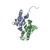


 PDBj
PDBj Assembly
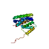

 Mass: 92.094 Da / Num. of mol.: 1 / Source method: obtained synthetically / Formula: C3H8O3
 Mass: 18.015 Da / Num. of mol.: 31 / Source method: isolated from a natural source / Formula: H2O
 Sample preparation
 Processing